 Movie
Movie Controller


+ Open data

- Basic information
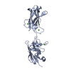
| Entry | Database: PDB / ID: 5ftx | |||||||||
|---|---|---|---|---|---|---|---|---|---|---|
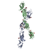
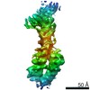
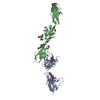
| Title | Structure of surface layer protein SbsC, domains 4-9 | |||||||||
 Components Components | SURFACE LAYER PROTEIN | |||||||||
 Keywords Keywords | CELL ADHESION / SURFACE LAYER / SELF-ASSEMBLY | |||||||||
| Function / homology |  Function and homology information Function and homology information | |||||||||
| Biological species |   GEOBACILLUS STEAROTHERMOPHILUS (bacteria) GEOBACILLUS STEAROTHERMOPHILUS (bacteria) | |||||||||
| Method |  X-RAY DIFFRACTION / SYNCHROTRON / MOLECULAR REPLACEMENT / Resolution: 4.1 Å X-RAY DIFFRACTION / SYNCHROTRON / MOLECULAR REPLACEMENT / Resolution: 4.1 Å | |||||||||
 Authors Authors | Dordic, A. / Pavkov-Keller, T. / Eder, M. / Egelseer, E.M. / Davis, K. / Mills, D. / Sleytr, U.B. / Kuehlbrandt, W. / Vonck, J. / Keller, W. | |||||||||
 Citation Citation | Journal: To be Published Title: Structure of Surface Layer Protein Sbsc Authors: Pavkov-Keller, T. / Dordic, A. / Eder, M. / Egelseer, E.M. / Davis, K. / Mills, D. / Sleytr, U.B. / Kuehlbrandt, W. / Vonck, J. / Keller, W. | |||||||||
| History |
|
- Structure visualization
| Structure viewer | Molecule: MolmilJmol/JSmol |
|---|
- Downloads & links
-Download
| PDBx/mmCIF format | 5ftx.cif.gz | 235.7 KB | Display | PDBx/mmCIF format |
|---|---|---|---|---|
| PDB format | pdb5ftx.ent.gz | 190.8 KB | Display | PDB format |
| PDBx/mmJSON format | 5ftx.json.gz | Tree view | PDBx/mmJSON format | |
| Others |  Other downloads Other downloads |
-Validation report
| Arichive directory | https://data.pdbj.org/pub/pdb/validation_reports/ft/5ftxftp://data.pdbj.org/pub/pdb/validation_reports/ft/5ftx | HTTPS FTP |
|---|
-Related structure data
| Related structure data | 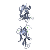 4uj8C  4uicS  4uieS 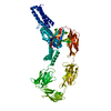 4uj6S 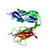 4uj7S S: Starting model for refinement C: citing same article ( |
|---|---|
| Similar structure data |


-Links
 PDBj
PDBj- Assembly
| Deposited unit | 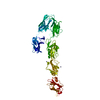
| ||||||||
|---|---|---|---|---|---|---|---|---|---|
| 1 |
| ||||||||
| Unit cell |
|
-Components
| #1: Protein | Mass: 67143.617 Da / Num. of mol.: 1 / Fragment: RSBSC, UNP RESIDUES 447-1099 Source method: isolated from a genetically manipulated source Details: STRUCTURE OF SURFACE LAYER PROTEIN SBSC, DOMAINS 4-9 Source: (gene. exp.) GEOBACILLUS STEAROTHERMOPHILUS (bacteria)Production host: | ||
|---|---|---|---|
| #2: Chemical | ChemComp-CA /   Mass: 40.078 Da / Num. of mol.: 5 / Source method: obtained synthetically / Formula: Ca Mass: 40.078 Da / Num. of mol.: 5 / Source method: obtained synthetically / Formula: Ca#3: Chemical |   Mass: 65.409 Da / Num. of mol.: 2 / Source method: obtained synthetically / Formula: Zn Mass: 65.409 Da / Num. of mol.: 2 / Source method: obtained synthetically / Formula: Zn |
-Experimental details
-Experiment
| Experiment | Method: X-RAY DIFFRACTION / Number of used crystals: 1 |
|---|
- Sample preparation
| Crystal | Density Matthews: 3.5 Å3/Da / Density % sol: 65 % / Description: DATA VERY ANISOTROPIC. |
|---|
-Data collection
| Diffraction | Mean temperature: 100 K |
|---|---|
| Diffraction source | Source: SYNCHROTRON / Site: ELETTRA  / Beamline: 5.2R / Wavelength: 1.14 / Beamline: 5.2R / Wavelength: 1.14 |
| Detector | Type: DECTRIS PILATUS 2M / Detector: PIXEL |
| Radiation | Protocol: SINGLE WAVELENGTH / Monochromatic (M) / Laue (L): M / Scattering type: x-ray |
| Radiation wavelength | Wavelength: 1.14 Å / Relative weight: 1 |
| Reflection | Resolution: 3.46→42.5 Å / Num. obs: 11056 / % possible obs: 87.6 % / Observed criterion σ(I): 3.6 / Redundancy: 2.9 % / Biso Wilson estimate: 87.26 Å2 / Rmerge(I) obs: 0.15 / Net I/σ(I): 6 |
| Reflection shell | Resolution: 3.46→3.66 Å / Redundancy: 2.2 % / Rmerge(I) obs: 0.15 / Mean I/σ(I) obs: 3.6 / % possible all: 72.6 |
- Processing
| Software |
| ||||||||||||||||||||||||||||
|---|---|---|---|---|---|---|---|---|---|---|---|---|---|---|---|---|---|---|---|---|---|---|---|---|---|---|---|---|---|
| Refinement | Method to determine structure: MOLECULAR REPLACEMENT Starting model: PDB ENTRIES 4UJ6, 4UIC, 4UIE AND 4UJ7 Resolution: 4.1→33.038 Å / SU ML: 0.45 / σ(F): 1.99 / Phase error: 28.84 / Stereochemistry target values: ML Details: DATA ANISOTROPIC, DATA WERE TRUNCATED TO 4.1 A FOR THE REFINEMENT. FOR SOME DOMAINS, SIDE CHAINS WERE POORLY DEFINED OR NOT VISIBLE.
| ||||||||||||||||||||||||||||
| Solvent computation | Shrinkage radii: 0.9 Å / VDW probe radii: 1.11 Å / Solvent model: FLAT BULK SOLVENT MODEL | ||||||||||||||||||||||||||||
| Refinement step | Cycle: LAST / Resolution: 4.1→33.038 Å
| ||||||||||||||||||||||||||||
| Refine LS restraints |
| ||||||||||||||||||||||||||||
| LS refinement shell |
|
