 Movie
Movie Controller

[English] 日本語
 Yorodumi
- PDB-5fsu: Crystal structure of Trypanosoma brucei macrodomain (crystal form 1) -
+ Open data

- Basic information
| Entry | Database: PDB / ID: 5fsu | ||||||
|---|---|---|---|---|---|---|---|
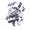
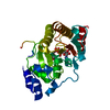
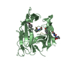
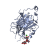
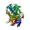
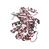
| Title | Crystal structure of Trypanosoma brucei macrodomain (crystal form 1) | ||||||
 Components Components | MACRODOMAIN | ||||||
 Keywords Keywords | HYDROLASE / MACRODOMAIN / ADP-RIBOSE BINDING | ||||||
| Function / homology |  Function and homology information Function and homology information | ||||||
| Biological species |  | ||||||
| Method |  X-RAY DIFFRACTION / SYNCHROTRON / MOLECULAR REPLACEMENT / Resolution: 1.95 Å X-RAY DIFFRACTION / SYNCHROTRON / MOLECULAR REPLACEMENT / Resolution: 1.95 Å | ||||||
 Authors Authors | Haikarainen, T. / Lehtio, L. | ||||||
 Citation Citation | Journal: Sci.Rep. / Year: 2016 Title: Proximal Adp-Ribose Hydrolysis in Trypanosomatids is Catalyzed by a Macrodomain. Authors: Haikarainen, T. / Lehtio, L. | ||||||
| History |
|
- Structure visualization
| Structure viewer | Molecule: MolmilJmol/JSmol |
|---|
- Downloads & links
-Download
| PDBx/mmCIF format | 5fsu.cif.gz | 106.2 KB | Display | PDBx/mmCIF format |
|---|---|---|---|---|
| PDB format | pdb5fsu.ent.gz | 82.7 KB | Display | PDB format |
| PDBx/mmJSON format | 5fsu.json.gz | Tree view | PDBx/mmJSON format | |
| Others |  Other downloads Other downloads |
-Validation report
| Summary document | 5fsu_validation.pdf.gz | 427 KB | Display | wwPDB validaton report |
|---|---|---|---|---|
| Full document | 5fsu_full_validation.pdf.gz | 429.7 KB | Display | |
| Data in XML | 5fsu_validation.xml.gz | 19.7 KB | Display | |
| Data in CIF | 5fsu_validation.cif.gz | 28.6 KB | Display | |
| Arichive directory | https://data.pdbj.org/pub/pdb/validation_reports/fs/5fsuftp://data.pdbj.org/pub/pdb/validation_reports/fs/5fsu | HTTPS FTP |
-Related structure data
| Related structure data |  5fsvC  5fsxC  5fsyC  5fszC 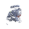 4iqyS C: citing same article ( S: Starting model for refinement |
|---|---|
| Similar structure data |


-Links
 PDBj
PDBj- Assembly
| Deposited unit | 
| ||||||||
|---|---|---|---|---|---|---|---|---|---|
| 1 | 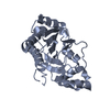
| ||||||||
| 2 | 
| ||||||||
| Unit cell |
|
-Components
| #1: Protein | Mass: 29175.555 Da / Num. of mol.: 2 Source method: isolated from a genetically manipulated source Source: (gene. exp.)  #2: Water | ChemComp-HOH / |  Mass: 18.015 Da / Num. of mol.: 213 / Source method: isolated from a natural source / Formula: H2O Mass: 18.015 Da / Num. of mol.: 213 / Source method: isolated from a natural source / Formula: H2O |
|---|
-Experimental details
-Experiment
| Experiment | Method: X-RAY DIFFRACTION / Number of used crystals: 1 |
|---|
- Sample preparation
| Crystal | Density Matthews: 3.16 Å3/Da / Density % sol: 61 % / Description: NONE |
|---|---|
| Crystal grow | pH: 7.5 Details: 0.2 M SODIUM SULFATE, 0.1 M BIS-TRIS PROPANE PH 7.5, 20 % W/V PEG 3350 |
-Data collection
| Diffraction | Mean temperature: 100 K |
|---|---|
| Diffraction source | Source: SYNCHROTRON / Site: Diamond  / Beamline: I03 / Wavelength: 0.97625 / Beamline: I03 / Wavelength: 0.97625 |
| Detector | Type: DECTRIS PIXEL / Detector: PIXEL / Date: May 3, 2015 / Details: MIRRORS |
| Radiation | Monochromator: DOUBLE CRYSTAL / Protocol: SINGLE WAVELENGTH / Monochromatic (M) / Laue (L): M / Scattering type: x-ray |
| Radiation wavelength | Wavelength: 0.97625 Å / Relative weight: 1 |
| Reflection | Resolution: 1.95→30 Å / Num. obs: 56483 / % possible obs: 99.7 % / Observed criterion σ(I): -3 / Redundancy: 15.1 % / Rmerge(I) obs: 0.09 / Net I/σ(I): 16.9 |
| Reflection shell | Resolution: 1.95→2 Å / Redundancy: 15.1 % / Mean I/σ(I) obs: 1.8 / % possible all: 99.7 |
- Processing
| Software |
| ||||||||||||||||||||||||||||||||||||||||||||||||||||||||||||||||||||||||||||||||||||||||||||||||||||||||||||||||||||||||||||||||||||||||||||||||||||||||||||||||||||||||||||||||||||||
|---|---|---|---|---|---|---|---|---|---|---|---|---|---|---|---|---|---|---|---|---|---|---|---|---|---|---|---|---|---|---|---|---|---|---|---|---|---|---|---|---|---|---|---|---|---|---|---|---|---|---|---|---|---|---|---|---|---|---|---|---|---|---|---|---|---|---|---|---|---|---|---|---|---|---|---|---|---|---|---|---|---|---|---|---|---|---|---|---|---|---|---|---|---|---|---|---|---|---|---|---|---|---|---|---|---|---|---|---|---|---|---|---|---|---|---|---|---|---|---|---|---|---|---|---|---|---|---|---|---|---|---|---|---|---|---|---|---|---|---|---|---|---|---|---|---|---|---|---|---|---|---|---|---|---|---|---|---|---|---|---|---|---|---|---|---|---|---|---|---|---|---|---|---|---|---|---|---|---|---|---|---|---|---|
| Refinement | Method to determine structure: MOLECULAR REPLACEMENT Starting model: PDB ENTRY 4IQY Resolution: 1.95→19.75 Å / Cor.coef. Fo:Fc: 0.971 / Cor.coef. Fo:Fc free: 0.958 / SU B: 3.992 / SU ML: 0.105 / Cross valid method: THROUGHOUT / ESU R: 0.12 / ESU R Free: 0.117 / Stereochemistry target values: MAXIMUM LIKELIHOOD / Details: HYDROGENS HAVE BEEN ADDED IN THE RIDING POSITIONS.
| ||||||||||||||||||||||||||||||||||||||||||||||||||||||||||||||||||||||||||||||||||||||||||||||||||||||||||||||||||||||||||||||||||||||||||||||||||||||||||||||||||||||||||||||||||||||
| Solvent computation | Ion probe radii: 0.8 Å / Shrinkage radii: 0.8 Å / VDW probe radii: 1.2 Å / Solvent model: MASK | ||||||||||||||||||||||||||||||||||||||||||||||||||||||||||||||||||||||||||||||||||||||||||||||||||||||||||||||||||||||||||||||||||||||||||||||||||||||||||||||||||||||||||||||||||||||
| Displacement parameters | Biso mean: 53.727 Å2
| ||||||||||||||||||||||||||||||||||||||||||||||||||||||||||||||||||||||||||||||||||||||||||||||||||||||||||||||||||||||||||||||||||||||||||||||||||||||||||||||||||||||||||||||||||||||
| Refinement step | Cycle: LAST / Resolution: 1.95→19.75 Å
| ||||||||||||||||||||||||||||||||||||||||||||||||||||||||||||||||||||||||||||||||||||||||||||||||||||||||||||||||||||||||||||||||||||||||||||||||||||||||||||||||||||||||||||||||||||||
| Refine LS restraints |
|