 Movie
Movie Controller

[English] 日本語
 Yorodumi
- PDB-5fkw: cryo-EM structure of the E. coli replicative DNA polymerase compl... -
+ Open data

- Basic information
| Entry | Database: PDB / ID: 5fkw | ||||||
|---|---|---|---|---|---|---|---|
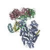
| Title | cryo-EM structure of the E. coli replicative DNA polymerase complex bound to DNA (DNA polymerase III alpha, beta, epsilon) | ||||||
 Components Components |
| ||||||
 Keywords Keywords | TRANSFERASE / DNA REPLICATION / DNA POLYMERASE III ALPHA / DNA POLYMERASE III BETA / DNA POLYMERASE III EPSILON | ||||||
| Function / homology |  Function and homology information Function and homology informationDNA polymerase III, core complex / Hda-beta clamp complex / bacterial-type DNA replication / replication inhibiting complex / DNA replication proofreading / DNA polymerase III complex / lagging strand elongation / replisome / regulation of DNA-templated DNA replication initiation / exonuclease activity ...DNA polymerase III, core complex / Hda-beta clamp complex / bacterial-type DNA replication / replication inhibiting complex / DNA replication proofreading / DNA polymerase III complex / lagging strand elongation / replisome / regulation of DNA-templated DNA replication initiation / exonuclease activity / DNA strand elongation involved in DNA replication / leading strand elongation / error-prone translesion synthesis / 3'-5' exonuclease activity / negative regulation of DNA-templated DNA replication initiation / DNA-templated DNA replication / DNA-directed DNA polymerase / DNA-directed DNA polymerase activity / DNA damage response / protein homodimerization activity / DNA binding / metal ion binding / identical protein binding / cytoplasm / cytosol Similarity search - Function | ||||||
| Biological species |  SYNTHETIC CONSTRUCT (others) | ||||||
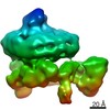
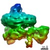
| Method | ELECTRON MICROSCOPY / single particle reconstruction / cryo EM / Resolution: 7.3 Å | ||||||
 Authors Authors | Fernandez-Leiro, R. / Conrad, J. / Scheres, S.H.W. / Lamers, M.H. | ||||||
 Citation Citation | Journal: Elife / Year: 2015 Title: cryo-EM structures of the replicative DNA polymerase reveal its dynamic interactions with the DNA sliding clamp, exonuclease and . Authors: Rafael Fernandez-Leiro / Julian Conrad / Sjors Hw Scheres / Meindert H Lamers /  Abstract: The replicative DNA polymerase PolIIIα from is a uniquely fast and processive enzyme. For its activity it relies on the DNA sliding clamp β, the proofreading exonuclease ε and the C-terminal ...The replicative DNA polymerase PolIIIα from is a uniquely fast and processive enzyme. For its activity it relies on the DNA sliding clamp β, the proofreading exonuclease ε and the C-terminal domain of the clamp loader subunit τ. Due to the dynamic nature of the four-protein complex it has long been refractory to structural characterization. Here we present the 8 Å resolution cryo-electron microscopy structures of DNA-bound and DNA-free states of the PolIII-clamp-exonuclease-τ complex. The structures show how the polymerase is tethered to the DNA through multiple contacts with the clamp and exonuclease. A novel contact between the polymerase and clamp is made in the DNA bound state, facilitated by a large movement of the polymerase tail domain and τ. These structures provide crucial insights into the organization of the catalytic core of the replisome and form an important step towards determining the structure of the complete holoenzyme. | ||||||
| History |
|
- Structure visualization
| Movie |
Movie viewer |
|---|---|
| Structure viewer | Molecule: MolmilJmol/JSmol |
- Downloads & links
-Download
| PDBx/mmCIF format | 5fkw.cif.gz | 354.5 KB | Display | PDBx/mmCIF format |
|---|---|---|---|---|
| PDB format | pdb5fkw.ent.gz | 271.1 KB | Display | PDB format |
| PDBx/mmJSON format | 5fkw.json.gz | Tree view | PDBx/mmJSON format | |
| Others |  Other downloads Other downloads |
-Validation report
| Arichive directory | https://data.pdbj.org/pub/pdb/validation_reports/fk/5fkwftp://data.pdbj.org/pub/pdb/validation_reports/fk/5fkw | HTTPS FTP |
|---|
-Related structure data
| Related structure data | 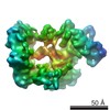 3202MC 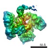 3198C 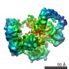 3201C 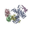 5fkuC 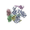 5fkvC C: citing same article ( M: map data used to model this data |
|---|---|
| Similar structure data |


-Links
 PDBj
PDBj


- Assembly
| Deposited unit | 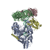
|
|---|---|
| 1 |
|
-Components
| #1: Protein | Mass: 130088.430 Da / Num. of mol.: 1 / Mutation: YES Source method: isolated from a genetically manipulated source Source: (gene. exp.) | ||||||
|---|---|---|---|---|---|---|---|
| #2: Protein | Mass: 40630.508 Da / Num. of mol.: 2 Source method: isolated from a genetically manipulated source Source: (gene. exp.) #3: Protein | | Mass: 27118.984 Da / Num. of mol.: 1 / Mutation: YES Source method: isolated from a genetically manipulated source Source: (gene. exp.) #4: DNA chain | | Mass: 7797.048 Da / Num. of mol.: 1 / Source method: obtained synthetically Details: TEMPLATE STRAND TCAGGAGTCCTTCGTCCTAGTACTACTCC PRIMER STRAND GGAGTAGTACTAGGACGAAGGACTC Source: (synth.) SYNTHETIC CONSTRUCT (others) #5: DNA chain | | Mass: 8796.659 Da / Num. of mol.: 1 / Source method: obtained synthetically Details: TEMPLATE STRAND TCAGGAGTCCTTCGTCCTAGTACTACTCC PRIMER STRAND GGAGTAGTACTAGGACGAAGGACTC Source: (synth.) SYNTHETIC CONSTRUCT (others) |
-Experimental details
-Experiment
| Experiment | Method: ELECTRON MICROSCOPY |
|---|---|
| EM experiment | Aggregation state: PARTICLE / 3D reconstruction method: single particle reconstruction |
- Sample preparation
| Component | Name: DNA POLYMERASE III CATALYTIC COMPLEX (ALPHA, EPSILON, BETA, TAU) Type: COMPLEX |
|---|---|
| Buffer solution | Name: 25 MM HEPES PH 7.5, 150 MM NACL, AND 2 MM DTT / pH: 7.5 / Details: 25 MM HEPES PH 7.5, 150 MM NACL, AND 2 MM DTT |
| Specimen | Conc.: 1 mg/ml / Embedding applied: NO / Shadowing applied: NO / Staining applied: NO / Vitrification applied: YES |
| Specimen support | Details: HOLEY CARBON |
| Vitrification | Instrument: FEI VITROBOT MARK III / Cryogen name: ETHANE / Details: LIQUID ETHANE |
- Electron microscopy imaging
| Experimental equipment |  Model: Titan Krios / Image courtesy: FEI Company |
|---|---|
| Microscopy | Model: FEI TITAN KRIOS / Date: May 12, 2014 |
| Electron gun | Electron source:  FIELD EMISSION GUN / Accelerating voltage: 300 kV / Illumination mode: FLOOD BEAM FIELD EMISSION GUN / Accelerating voltage: 300 kV / Illumination mode: FLOOD BEAM |
| Electron lens | Mode: BRIGHT FIELD / Nominal magnification: 64000 X / Calibrated magnification: 28409 X / Nominal defocus max: 4000 nm / Nominal defocus min: 2000 nm / Cs: 2.7 mm |
| Specimen holder | Temperature: 85 K |
| Image recording | Electron dose: 40 e/Å2 / Film or detector model: GATAN K2 QUANTUM (4k x 4k) |
| Image scans | Num. digital images: 1350 |
- Processing
| Symmetry | Point symmetry: C1 (asymmetric) | ||||||||||||
|---|---|---|---|---|---|---|---|---|---|---|---|---|---|
| 3D reconstruction | Resolution: 7.3 Å / Resolution method: FSC 0.143 CUT-OFF / Num. of particles: 40582 / Refinement type: HALF-MAPS REFINED INDEPENDENTLY / Symmetry type: POINT | ||||||||||||
| Refinement | Highest resolution: 7.3 Å | ||||||||||||
| Refinement step | Cycle: LAST / Highest resolution: 7.3 Å
|
