 Movie
Movie Controller

[English] 日本語
 Yorodumi
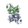
- PDB-5fi9: Closed form of murine Acid Sphingomyelinase in complex with bisph... -
+ Open data

- Basic information
| Entry | Database: PDB / ID: 5fi9 | |||||||||
|---|---|---|---|---|---|---|---|---|---|---|
| Title | Closed form of murine Acid Sphingomyelinase in complex with bisphosphonate inhibitor AbPA | |||||||||
 Components Components | Sphingomyelin phosphodiesterase | |||||||||
 Keywords Keywords | HYDROLASE / SMPD1 / ASM / ASMase / saposin | |||||||||
| Function / homology |  Function and homology information Function and homology informationacid sphingomyelin phosphodiesterase activity / sphingomyelin catabolic process / sphingomyelin phosphodiesterase / Glycosphingolipid catabolism / sphingomyelin phosphodiesterase activity / phospholipase C / phosphatidylcholine phospholipase C activity / endolysosome / lamellar body / termination of signal transduction ...acid sphingomyelin phosphodiesterase activity / sphingomyelin catabolic process / sphingomyelin phosphodiesterase / Glycosphingolipid catabolism / sphingomyelin phosphodiesterase activity / phospholipase C / phosphatidylcholine phospholipase C activity / endolysosome / lamellar body / termination of signal transduction / ceramide metabolic process / hydrolase activity, acting on glycosyl bonds / response to type I interferon / plasma membrane repair / ceramide biosynthetic process / cholesterol efflux / response to ionizing radiation / response to tumor necrosis factor / pyroptotic inflammatory response / positive regulation of endocytosis / cholesterol metabolic process / negative regulation of MAPK cascade / lipid droplet / cellular response to calcium ion / response to interleukin-1 / response to cocaine / wound healing / response to virus / cellular response to UV / positive regulation of viral entry into host cell / lysosome / endosome / positive regulation of apoptotic process / response to xenobiotic stimulus / symbiont entry into host cell / : / zinc ion binding / plasma membrane Similarity search - Function | |||||||||
| Biological species |  | |||||||||
| Method |  X-RAY DIFFRACTION / SYNCHROTRON / MOLECULAR REPLACEMENT / Resolution: 2.543 Å X-RAY DIFFRACTION / SYNCHROTRON / MOLECULAR REPLACEMENT / Resolution: 2.543 Å | |||||||||
 Authors Authors | Gorelik, A. / Illes, K. / Heinz, L.X. / Superti-Furga, G. / Nagar, B. | |||||||||
 Citation Citation | Journal: Nat Commun / Year: 2016 Title: Crystal structure of mammalian acid sphingomyelinase. Authors: Gorelik, A. / Illes, K. / Heinz, L.X. / Superti-Furga, G. / Nagar, B. | |||||||||
| History |
|
- Structure visualization
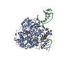
| Structure viewer | Molecule: MolmilJmol/JSmol |
|---|
- Downloads & links
-Download
| PDBx/mmCIF format | 5fi9.cif.gz | 408.4 KB | Display | PDBx/mmCIF format |
|---|---|---|---|---|
| PDB format | pdb5fi9.ent.gz | 341.5 KB | Display | PDB format |
| PDBx/mmJSON format | 5fi9.json.gz | Tree view | PDBx/mmJSON format | |
| Others |  Other downloads Other downloads |
-Validation report
| Arichive directory | https://data.pdbj.org/pub/pdb/validation_reports/fi/5fi9ftp://data.pdbj.org/pub/pdb/validation_reports/fi/5fi9 | HTTPS FTP |
|---|
-Related structure data
| Related structure data |  5fibC  5ficC  5hqnC 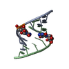 5hnqS C: citing same article ( S: Starting model for refinement |
|---|---|
| Similar structure data |


-Links
 PDBj
PDBj
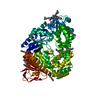
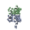
- Assembly
| Deposited unit | 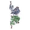
| ||||||||
|---|---|---|---|---|---|---|---|---|---|
| 1 | 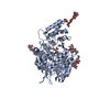
| ||||||||
| 2 | 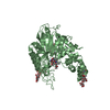
| ||||||||
| Unit cell |
|
-Components
-Protein , 1 types, 2 molecules AB
| #1: Protein | Mass: 60431.633 Da / Num. of mol.: 2 / Fragment: UNP residues 84-611 Source method: isolated from a genetically manipulated source Source: (gene. exp.)   Spodoptera frugiperda (fall armyworm) Spodoptera frugiperda (fall armyworm)References: UniProt: Q04519, sphingomyelin phosphodiesterase |
|---|
-Sugars , 5 types, 9 molecules
| #2: Polysaccharide | 2-acetamido-2-deoxy-beta-D-glucopyranose-(1-4)-2-acetamido-2-deoxy-beta-D-glucopyranose Source method: isolated from a genetically manipulated source #3: Polysaccharide | Source method: isolated from a genetically manipulated source #4: Polysaccharide | alpha-D-mannopyranose-(1-3)-[alpha-D-mannopyranose-(1-6)]beta-D-mannopyranose-(1-4)-2-acetamido-2- ...alpha-D-mannopyranose-(1-3)-[alpha-D-mannopyranose-(1-6)]beta-D-mannopyranose-(1-4)-2-acetamido-2-deoxy-beta-D-glucopyranose-(1-4)-[alpha-L-fucopyranose-(1-6)]2-acetamido-2-deoxy-beta-D-glucopyranose | Source method: isolated from a genetically manipulated source #5: Polysaccharide | beta-D-mannopyranose-(1-4)-2-acetamido-2-deoxy-beta-D-glucopyranose-(1-4)-[alpha-L-fucopyranose-(1- ...beta-D-mannopyranose-(1-4)-2-acetamido-2-deoxy-beta-D-glucopyranose-(1-4)-[alpha-L-fucopyranose-(1-6)]2-acetamido-2-deoxy-beta-D-glucopyranose | Source method: isolated from a genetically manipulated source #6: Polysaccharide | alpha-D-mannopyranose-(1-6)-beta-D-mannopyranose-(1-4)-2-acetamido-2-deoxy-beta-D-glucopyranose-(1- ...alpha-D-mannopyranose-(1-6)-beta-D-mannopyranose-(1-4)-2-acetamido-2-deoxy-beta-D-glucopyranose-(1-4)-[alpha-L-fucopyranose-(1-6)]2-acetamido-2-deoxy-beta-D-glucopyranose | Source method: isolated from a genetically manipulated source |
|---|
-Non-polymers , 3 types, 60 molecules 
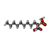

| #7: Chemical | ChemComp-ZN /  Mass: 65.409 Da / Num. of mol.: 4 / Source method: obtained synthetically / Formula: Zn Mass: 65.409 Da / Num. of mol.: 4 / Source method: obtained synthetically / Formula: Zn#8: Chemical |  Mass: 317.256 Da / Num. of mol.: 2 / Source method: obtained synthetically / Formula: C10H25NO6P2 Mass: 317.256 Da / Num. of mol.: 2 / Source method: obtained synthetically / Formula: C10H25NO6P2#9: Water | ChemComp-HOH / | Mass: 18.015 Da / Num. of mol.: 54 / Source method: isolated from a natural source / Formula: H2O |
|---|
-Details
| Has protein modification | Y |
|---|
-Experimental details
-Experiment
| Experiment | Method: X-RAY DIFFRACTION / Number of used crystals: 1 |
|---|
- Sample preparation
| Crystal | Density Matthews: 2.38 Å3/Da / Density % sol: 48.23 % |
|---|---|
| Crystal grow | Temperature: 293 K / Method: vapor diffusion, sitting drop / Details: lithium acetate, PEG 3350 |
-Data collection
| Diffraction | Mean temperature: 100 K |
|---|---|
| Diffraction source | Source: SYNCHROTRON / Site: CLSI  / Beamline: 08B1-1 / Wavelength: 1.28154 Å / Beamline: 08B1-1 / Wavelength: 1.28154 Å |
| Detector | Type: RAYONIX MX300HE / Detector: CCD / Date: May 20, 2015 |
| Radiation | Protocol: SINGLE WAVELENGTH / Monochromatic (M) / Laue (L): M / Scattering type: x-ray |
| Radiation wavelength | Wavelength: 1.28154 Å / Relative weight: 1 |
| Reflection | Resolution: 2.543→46.654 Å / Num. obs: 35624 / % possible obs: 97.2 % / Redundancy: 2.9 % / Net I/σ(I): 9.68 |
- Processing
| Software |
| |||||||||||||||||||||||||||||||||||||||||||||||||||||||||||||||||||||||||||||||||||||||||||
|---|---|---|---|---|---|---|---|---|---|---|---|---|---|---|---|---|---|---|---|---|---|---|---|---|---|---|---|---|---|---|---|---|---|---|---|---|---|---|---|---|---|---|---|---|---|---|---|---|---|---|---|---|---|---|---|---|---|---|---|---|---|---|---|---|---|---|---|---|---|---|---|---|---|---|---|---|---|---|---|---|---|---|---|---|---|---|---|---|---|---|---|---|
| Refinement | Method to determine structure: MOLECULAR REPLACEMENT Starting model: 5HNQ Resolution: 2.543→46.654 Å / SU ML: 0.36 / Cross valid method: FREE R-VALUE / σ(F): 1.77 / Phase error: 27.69 / Stereochemistry target values: ML
| |||||||||||||||||||||||||||||||||||||||||||||||||||||||||||||||||||||||||||||||||||||||||||
| Solvent computation | Shrinkage radii: 0.9 Å / VDW probe radii: 1.11 Å / Solvent model: FLAT BULK SOLVENT MODEL | |||||||||||||||||||||||||||||||||||||||||||||||||||||||||||||||||||||||||||||||||||||||||||
| Refinement step | Cycle: LAST / Resolution: 2.543→46.654 Å
| |||||||||||||||||||||||||||||||||||||||||||||||||||||||||||||||||||||||||||||||||||||||||||
| Refine LS restraints |
| |||||||||||||||||||||||||||||||||||||||||||||||||||||||||||||||||||||||||||||||||||||||||||
| LS refinement shell |
|
