 Movie
Movie Controller


+ Open data

- Basic information
| Entry | Database: PDB / ID: 5fax | |||||||||
|---|---|---|---|---|---|---|---|---|---|---|
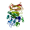
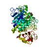
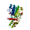
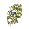
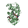
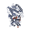
| Title | Structure of subtilase SubHal from Bacillus halmapalus | |||||||||
 Components Components | Subtilase SubHal from Bacillus halmapalus | |||||||||
 Keywords Keywords | HYDROLASE / Protease / Subtilase / Calcium binding | |||||||||
| Function / homology |  Function and homology information Function and homology information3.4.21.14 / serine-type endopeptidase activity / proteolysis / metal ion binding Similarity search - Function | |||||||||
| Biological species |  Bacillus halmapalus (bacteria) Bacillus halmapalus (bacteria) | |||||||||
| Method |  X-RAY DIFFRACTION / SYNCHROTRON / MOLECULAR REPLACEMENT / Resolution: 2 Å X-RAY DIFFRACTION / SYNCHROTRON / MOLECULAR REPLACEMENT / Resolution: 2 Å | |||||||||
 Authors Authors | Dohnalek, J. / Brzozowski, A.M. / Svendsen, A. / Wilson, K.S. | |||||||||
 Citation Citation | Book title: Understanding enzymes; Function, Design, Engineering and Analysis Journal: Book / Year: 2016 Title: Stabilization of Enzymes by Metal Binding: Structures of Two Alkalophilic Bacillus Subtilases and Analysis of the Second Metal-Binding Site of the Subtilase Family Authors: Dohnalek, J. / McAuley, K.E. / Brzozowski, A.M. / Oestergaard, P.R. / Svendsen, A. / Wilson, K.S. | |||||||||
| History |
|
- Structure visualization
| Structure viewer | Molecule: MolmilJmol/JSmol |
|---|
- Downloads & links
-Download
| PDBx/mmCIF format | 5fax.cif.gz | 187 KB | Display | PDBx/mmCIF format |
|---|---|---|---|---|
| PDB format | pdb5fax.ent.gz | 145.2 KB | Display | PDB format |
| PDBx/mmJSON format | 5fax.json.gz | Tree view | PDBx/mmJSON format | |
| Others |  Other downloads Other downloads |
-Validation report
| Arichive directory | https://data.pdbj.org/pub/pdb/validation_reports/fa/5faxftp://data.pdbj.org/pub/pdb/validation_reports/fa/5fax | HTTPS FTP |
|---|
-Related structure data
| Related structure data |  5fbzSC  5ffnC S: Starting model for refinement C: citing same article ( |
|---|---|
| Similar structure data |


-Links
 PDBj
PDBj

- Assembly
| Deposited unit | 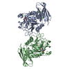
| ||||||||
|---|---|---|---|---|---|---|---|---|---|
| 1 | 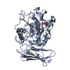
| ||||||||
| 2 | 
| ||||||||
| Unit cell |
|
-Components
| #1: Protein | Mass: 45356.980 Da / Num. of mol.: 2 Source method: isolated from a genetically manipulated source Details: N-terminal asparagine is naturally modified to N-carboxyasparagine. There is no associated Uniprot entry, however the sequence is available in patent WO 2004083362 Source: (gene. exp.) Bacillus halmapalus (bacteria) / Production host: #2: Chemical | ChemComp-CA /   Mass: 40.078 Da / Num. of mol.: 6 / Source method: obtained synthetically / Formula: Ca Mass: 40.078 Da / Num. of mol.: 6 / Source method: obtained synthetically / Formula: Ca#3: Water | ChemComp-HOH / |  Mass: 18.015 Da / Num. of mol.: 662 / Source method: isolated from a natural source / Formula: H2O Mass: 18.015 Da / Num. of mol.: 662 / Source method: isolated from a natural source / Formula: H2O |
|---|
-Experimental details
-Experiment
| Experiment | Method: X-RAY DIFFRACTION / Number of used crystals: 1 |
|---|
- Sample preparation
| Crystal | Density Matthews: 2.36 Å3/Da / Density % sol: 47.8 % / Description: Elongated plates |
|---|---|
| Crystal grow | Temperature: 291 K / Method: vapor diffusion, hanging drop / pH: 6.5 Details: Protein concentration 24 mg ml-1 in 50 mM sodium cacodylate buffer, pH 6.5, 2 mM CaCl2 and 100 mM NaCl. The enzyme was crystallised by hanging drop vapour diffusion in drops containing 1 ...Details: Protein concentration 24 mg ml-1 in 50 mM sodium cacodylate buffer, pH 6.5, 2 mM CaCl2 and 100 mM NaCl. The enzyme was crystallised by hanging drop vapour diffusion in drops containing 1 microliter of protein and 1 microliter of reservoir solution over 0.5 ml of reservoir: Hampton Screen 2 no. 23 (10% dioxane, 0.1 M 2-(N-Morpholino)-ethanesulphonic acid (MES) buffer, pH 6.5, 1.6 M ammonium sulphate). Clusters of elongated plates appeared after several days. |
-Data collection
| Diffraction | Mean temperature: 100 K |
|---|---|
| Diffraction source | Source: SYNCHROTRON / Site: ESRF  / Beamline: ID14-2 / Wavelength: 0.933 Å / Beamline: ID14-2 / Wavelength: 0.933 Å |
| Detector | Type: ADSC QUANTUM 4 / Detector: CCD / Date: Mar 1, 2002 |
| Radiation | Protocol: SINGLE WAVELENGTH / Monochromatic (M) / Laue (L): M / Scattering type: x-ray |
| Radiation wavelength | Wavelength: 0.933 Å / Relative weight: 1 |
| Reflection | Resolution: 2→30 Å / Num. all: 41891 / Num. obs: 41891 / % possible obs: 74 % / Observed criterion σ(I): -3 / Redundancy: 4.3 % / Biso Wilson estimate: 13.2 Å2 / Rmerge(I) obs: 0.101 / Net I/σ(I): 6 |
| Reflection shell | Resolution: 2→2.05 Å / Rmerge(I) obs: 0.268 / Mean I/σ(I) obs: 2.4 / % possible all: 49.1 |
- Processing
| Software |
| ||||||||||||||||||||||||||||||||||||||||||||||||||||||||||||||||||||||||||||||||||||||||||||||||||||||||||||||||||||||||||||||||||||||||||||||||||||||||||||||||||||||||||||||||||||||
|---|---|---|---|---|---|---|---|---|---|---|---|---|---|---|---|---|---|---|---|---|---|---|---|---|---|---|---|---|---|---|---|---|---|---|---|---|---|---|---|---|---|---|---|---|---|---|---|---|---|---|---|---|---|---|---|---|---|---|---|---|---|---|---|---|---|---|---|---|---|---|---|---|---|---|---|---|---|---|---|---|---|---|---|---|---|---|---|---|---|---|---|---|---|---|---|---|---|---|---|---|---|---|---|---|---|---|---|---|---|---|---|---|---|---|---|---|---|---|---|---|---|---|---|---|---|---|---|---|---|---|---|---|---|---|---|---|---|---|---|---|---|---|---|---|---|---|---|---|---|---|---|---|---|---|---|---|---|---|---|---|---|---|---|---|---|---|---|---|---|---|---|---|---|---|---|---|---|---|---|---|---|---|---|
| Refinement | Method to determine structure: MOLECULAR REPLACEMENT Starting model: Complex of SubHal with CI2A inhibitor, PDB ID 5FBZ Resolution: 2→30 Å / Cor.coef. Fo:Fc: 0.888 / SU B: 3.193 / SU ML: 0.097 / Cross valid method: FREE R-VALUE / σ(F): 0 / ESU R: 0.342 / ESU R Free: 0.287 / Stereochemistry target values: MAXIMUM LIKELIHOOD / Details: HYDROGENS HAVE BEEN ADDED IN THE RIDING POSITIONS
| ||||||||||||||||||||||||||||||||||||||||||||||||||||||||||||||||||||||||||||||||||||||||||||||||||||||||||||||||||||||||||||||||||||||||||||||||||||||||||||||||||||||||||||||||||||||
| Solvent computation | Ion probe radii: 0.8 Å / Shrinkage radii: 0.8 Å / VDW probe radii: 1.4 Å / Solvent model: BABINET MODEL WITH MASK | ||||||||||||||||||||||||||||||||||||||||||||||||||||||||||||||||||||||||||||||||||||||||||||||||||||||||||||||||||||||||||||||||||||||||||||||||||||||||||||||||||||||||||||||||||||||
| Displacement parameters | Biso mean: 14.77 Å2
| ||||||||||||||||||||||||||||||||||||||||||||||||||||||||||||||||||||||||||||||||||||||||||||||||||||||||||||||||||||||||||||||||||||||||||||||||||||||||||||||||||||||||||||||||||||||
| Refinement step | Cycle: 1 / Resolution: 2→30 Å
| ||||||||||||||||||||||||||||||||||||||||||||||||||||||||||||||||||||||||||||||||||||||||||||||||||||||||||||||||||||||||||||||||||||||||||||||||||||||||||||||||||||||||||||||||||||||
| Refine LS restraints |
|