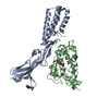
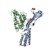
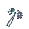
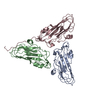
Acrosome Reaction and Sperm:Oocyte Membrane Binding / protein complex involved in cell-cell adhesion / syncytium formation by cell-cell fusion / sperm-egg recognition / protein binding involved in heterotypic cell-cell adhesion / fusion of sperm to egg plasma membrane involved in single fertilization / acrosomal membrane / Post-translational modification: synthesis of GPI-anchored proteins / binding of sperm to zona pellucida / microvillus membrane ...Acrosome Reaction and Sperm:Oocyte Membrane Binding / protein complex involved in cell-cell adhesion / syncytium formation by cell-cell fusion / sperm-egg recognition / protein binding involved in heterotypic cell-cell adhesion / fusion of sperm to egg plasma membrane involved in single fertilization / acrosomal membrane / Post-translational modification: synthesis of GPI-anchored proteins / binding of sperm to zona pellucida / microvillus membrane / single fertilization / acrosomal vesicle / signaling receptor activity / cell adhesion / signaling receptor binding / external side of plasma membrane / endoplasmic reticulum membrane / protein homodimerization activity / extracellular region / membrane / identical protein binding / plasma membrane Similarity search - Function
 Movie
Movie Controller


 Open data
 Basic information
 Components
 Keywords
 Function and homology information
 Homo sapiens (human)
 X-RAY DIFFRACTION /
  Authors
 Citation
 Structure visualization
 Downloads & links
 Other downloads


 PDBj
PDBj Assembly


 Type: D-saccharide, beta linking / Mass: 221.208 Da / Num. of mol.: 1


 Mass: 35.453 Da / Num. of mol.: 1 / Source method: obtained synthetically / Formula: Cl
 Mass: 92.094 Da / Num. of mol.: 1 / Source method: obtained synthetically / Formula: C3H8O3
 Sample preparation
 / Beamline: 08ID-1 / Wavelength: 1.77 Å
 Processing