 Movie
Movie Controller

[English] 日本語
 Yorodumi
- PDB-5eul: Structure of the SecA-SecY complex with a translocating polypepti... -
+ Open data

- Basic information
| Entry | Database: PDB / ID: 5eul | ||||||
|---|---|---|---|---|---|---|---|
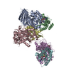
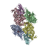
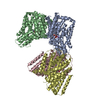
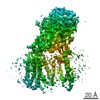
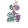
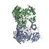
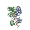
| Title | Structure of the SecA-SecY complex with a translocating polypeptide substrate | ||||||
 Components Components |
| ||||||
 Keywords Keywords | PROTEIN TRANSPORT / SecY / SecA / ATPase / channel | ||||||
| Function / homology |  Function and homology information Function and homology informationcell envelope Sec protein transport complex / protein-exporting ATPase activity / protein-secreting ATPase / protein transport by the Sec complex / intracellular protein transmembrane transport / protein import / protein secretion / transmembrane protein transporter activity / protein targeting / membrane raft ...cell envelope Sec protein transport complex / protein-exporting ATPase activity / protein-secreting ATPase / protein transport by the Sec complex / intracellular protein transmembrane transport / protein import / protein secretion / transmembrane protein transporter activity / protein targeting / membrane raft / ATP binding / metal ion binding / plasma membrane / cytoplasm Similarity search - Function | ||||||
| Biological species |  synthetic construct (others) Geobacillus thermodenitrificans (bacteria) | ||||||
| Method |  X-RAY DIFFRACTION / SYNCHROTRON / MAD / Resolution: 3.7 Å X-RAY DIFFRACTION / SYNCHROTRON / MAD / Resolution: 3.7 Å | ||||||
 Authors Authors | Li, L. / Park, E. / Ling, J. / Ingram, J. / Ploegh, H. / Rapoport, T.A. | ||||||
| Funding support |  United States, 1items United States, 1items
| ||||||
 Citation Citation | Journal: Nature / Year: 2016 Title: Crystal structure of a substrate-engaged SecY protein-translocation channel. Authors: Li, L. / Park, E. / Ling, J. / Ingram, J. / Ploegh, H. / Rapoport, T.A. | ||||||
| History |
|
- Structure visualization
| Structure viewer | Molecule: MolmilJmol/JSmol |
|---|
- Downloads & links
-Download
| PDBx/mmCIF format | 5eul.cif.gz | 284.1 KB | Display | PDBx/mmCIF format |
|---|---|---|---|---|
| PDB format | pdb5eul.ent.gz | 225.1 KB | Display | PDB format |
| PDBx/mmJSON format | 5eul.json.gz | Tree view | PDBx/mmJSON format | |
| Others |  Other downloads Other downloads |
-Validation report
| Arichive directory | https://data.pdbj.org/pub/pdb/validation_reports/eu/5eulftp://data.pdbj.org/pub/pdb/validation_reports/eu/5eul | HTTPS FTP |
|---|
-Related structure data
| Similar structure data |
|---|

-Links
 PDBj
PDBj


- Assembly
| Deposited unit | 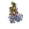
| ||||||||
|---|---|---|---|---|---|---|---|---|---|
| 1 |
| ||||||||
| Unit cell |
|
-Components
-Protein translocase subunit ... , 2 types, 2 molecules AY
| #1: Protein | Mass: 94360.195 Da / Num. of mol.: 1 Source method: isolated from a genetically manipulated source Source: (gene. exp.) Strain: 168 / Gene: secA, div+, BSU35300 / Production host: |
|---|---|
| #2: Protein | Mass: 46768.301 Da / Num. of mol.: 1 Source method: isolated from a genetically manipulated source Source: (gene. exp.) Geobacillus thermodenitrificans (strain NG80-2) (bacteria)Strain: NG80-2 / Gene: secY, GTNG_0125 / Production host: |
-Protein / Antibody , 2 types, 2 molecules EV
| #3: Protein | Mass: 8249.600 Da / Num. of mol.: 1 Source method: isolated from a genetically manipulated source Source: (gene. exp.) Geobacillus thermodenitrificans (strain NG80-2) (bacteria)Strain: NG80-2 / Gene: GTNG_0091 / Production host: |
|---|---|
| #4: Antibody | Mass: 14475.136 Da / Num. of mol.: 1 Source method: isolated from a genetically manipulated source Source: (gene. exp.) |
-Non-polymers , 4 types, 21 molecules 


| #5: Chemical | ChemComp-MG /  Mass: 24.305 Da / Num. of mol.: 1 / Source method: obtained synthetically / Formula: Mg Mass: 24.305 Da / Num. of mol.: 1 / Source method: obtained synthetically / Formula: Mg |
|---|---|
| #6: Chemical | ChemComp-BEF /  Mass: 66.007 Da / Num. of mol.: 1 / Source method: obtained synthetically / Formula: BeF3 Mass: 66.007 Da / Num. of mol.: 1 / Source method: obtained synthetically / Formula: BeF3 |
| #7: Chemical | ChemComp-ADP /  Mass: 427.201 Da / Num. of mol.: 1 / Source method: obtained synthetically / Formula: C10H15N5O10P2 / Comment: ADP, energy-carrying molecule*YM Mass: 427.201 Da / Num. of mol.: 1 / Source method: obtained synthetically / Formula: C10H15N5O10P2 / Comment: ADP, energy-carrying molecule*YM |
| #8: Chemical | ChemComp-TBR /  Mass: 2044.535 Da / Num. of mol.: 18 / Source method: obtained synthetically / Formula: Br12Ta6 Mass: 2044.535 Da / Num. of mol.: 18 / Source method: obtained synthetically / Formula: Br12Ta6 |
-Details
| Has protein modification | Y |
|---|
-Experimental details
-Experiment
| Experiment | Method: X-RAY DIFFRACTION / Number of used crystals: 1 |
|---|
- Sample preparation
| Crystal | Density Matthews: 3.89 Å3/Da / Density % sol: 68.35 % |
|---|---|
| Crystal grow | Temperature: 295 K / Method: vapor diffusion, hanging drop / pH: 8.5 Details: 21-24% polyethylene glycol 1500, 100mM Tris-HCl pH8.5, 50-100 mM MgAc2, 2% 2-methyl-2,4-pentandiol PH range: 8.5 |
-Data collection
| Diffraction | Mean temperature: 100 K |
|---|---|
| Diffraction source | Source: SYNCHROTRON / Site: APS / Beamline: 23-ID-D / Wavelength: 1.2782 Å |
| Detector | Type: DECTRIS PILATUS 6M / Detector: PIXEL / Date: Jun 18, 2015 |
| Radiation | Monochromator: Si(111) / Protocol: MAD / Monochromatic (M) / Laue (L): M / Scattering type: x-ray |
| Radiation wavelength | Wavelength: 1.2782 Å / Relative weight: 1 |
| Reflection | Resolution: 3.7→54 Å / Num. obs: 53847 / % possible obs: 99 % / Redundancy: 10.6 % / CC1/2: 0.998 / Rmerge(I) obs: 0.085 / Net I/σ(I): 9.3 |
| Reflection shell | Resolution: 3.7→3.8 Å / Redundancy: 10.2 % / Mean I/σ(I) obs: 0.4 / % possible all: 100 |
- Processing
| Software |
| |||||||||||||||||||||||||||||||||||||||||||||||||||||||||||||||||||||||||||||||||||||||||||||||||||||||||||||||||||||||||||||||||||||||||||||||||||
|---|---|---|---|---|---|---|---|---|---|---|---|---|---|---|---|---|---|---|---|---|---|---|---|---|---|---|---|---|---|---|---|---|---|---|---|---|---|---|---|---|---|---|---|---|---|---|---|---|---|---|---|---|---|---|---|---|---|---|---|---|---|---|---|---|---|---|---|---|---|---|---|---|---|---|---|---|---|---|---|---|---|---|---|---|---|---|---|---|---|---|---|---|---|---|---|---|---|---|---|---|---|---|---|---|---|---|---|---|---|---|---|---|---|---|---|---|---|---|---|---|---|---|---|---|---|---|---|---|---|---|---|---|---|---|---|---|---|---|---|---|---|---|---|---|---|---|---|---|
| Refinement | Method to determine structure: MAD / Resolution: 3.7→53.852 Å / SU ML: 0.71 / Cross valid method: FREE R-VALUE / σ(F): 1.91 / Phase error: 42.14 / Stereochemistry target values: ML
| |||||||||||||||||||||||||||||||||||||||||||||||||||||||||||||||||||||||||||||||||||||||||||||||||||||||||||||||||||||||||||||||||||||||||||||||||||
| Solvent computation | Shrinkage radii: 0.9 Å / VDW probe radii: 1.11 Å / Solvent model: FLAT BULK SOLVENT MODEL | |||||||||||||||||||||||||||||||||||||||||||||||||||||||||||||||||||||||||||||||||||||||||||||||||||||||||||||||||||||||||||||||||||||||||||||||||||
| Refinement step | Cycle: LAST / Resolution: 3.7→53.852 Å
| |||||||||||||||||||||||||||||||||||||||||||||||||||||||||||||||||||||||||||||||||||||||||||||||||||||||||||||||||||||||||||||||||||||||||||||||||||
| Refine LS restraints |
| |||||||||||||||||||||||||||||||||||||||||||||||||||||||||||||||||||||||||||||||||||||||||||||||||||||||||||||||||||||||||||||||||||||||||||||||||||
| LS refinement shell |
|
