 Movie
Movie Controller

[English] 日本語
 Yorodumi
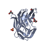
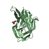
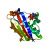
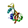
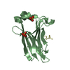
- PDB-5ete: Structure of pathogen-related yeast protein, Pry1 in complex with... -
+ Open data

- Basic information
| Entry | Database: PDB / ID: 5ete | ||||||
|---|---|---|---|---|---|---|---|
| Title | Structure of pathogen-related yeast protein, Pry1 in complex with a competitive inhibitor of cholesterol binding | ||||||
 Components Components | Pry1p | ||||||
 Keywords Keywords | Sterol binding protein / TAPs / testis specific proteins / Tpx / antigen 5 / Ag5 / pathogenesis related-1 / PR-1 / Sc7 / CAP / cysteine-rich secretory protein / CRISP | ||||||
| Function / homology | Pathogenesis-related Protein p14a / CAP / 3-Layer(aba) Sandwich / Alpha Beta / 1,4-DIETHYLENE DIOXIDE / :  Function and homology information Function and homology information | ||||||
| Biological species |  | ||||||
| Method |  X-RAY DIFFRACTION / MOLECULAR REPLACEMENT / Resolution: 2.1 Å X-RAY DIFFRACTION / MOLECULAR REPLACEMENT / Resolution: 2.1 Å | ||||||
 Authors Authors | Asojo, O.A. | ||||||
 Citation Citation | Journal: Sci Rep / Year: 2016 Title: Structural and functional characterization of the CAP domain of pathogen-related yeast 1 (Pry1) protein. Authors: Darwiche, R. / Kelleher, A. / Hudspeth, E.M. / Schneiter, R. / Asojo, O.A. | ||||||
| History |
|
- Structure visualization
| Structure viewer | Molecule: MolmilJmol/JSmol |
|---|
- Downloads & links
-Download
| PDBx/mmCIF format | 5ete.cif.gz | 66.7 KB | Display | PDBx/mmCIF format |
|---|---|---|---|---|
| PDB format | pdb5ete.ent.gz | 49.3 KB | Display | PDB format |
| PDBx/mmJSON format | 5ete.json.gz | Tree view | PDBx/mmJSON format | |
| Others |  Other downloads Other downloads |
-Validation report
| Arichive directory | https://data.pdbj.org/pub/pdb/validation_reports/et/5eteftp://data.pdbj.org/pub/pdb/validation_reports/et/5ete | HTTPS FTP |
|---|
-Related structure data
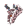
| Related structure data | 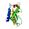 5jysC 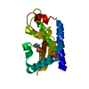 1smbS S: Starting model for refinement C: citing same article ( |
|---|---|
| Similar structure data |


-Links
 PDBj
PDBj- Assembly
| Deposited unit | 
| ||||||||||||||||||
|---|---|---|---|---|---|---|---|---|---|---|---|---|---|---|---|---|---|---|---|
| 1 |
| ||||||||||||||||||
| Unit cell |
| ||||||||||||||||||
| Components on special symmetry positions |
|
-Components
| #1: Protein | Mass: 15903.875 Da / Num. of mol.: 1 / Fragment: residues 158-306 Source method: isolated from a genetically manipulated source Source: (gene. exp.) Gene: PRY1, H819_YJM1434J00135 / Production host:  Pichia (fungus) / References: UniProt: A0A0C6AG41 Pichia (fungus) / References: UniProt: A0A0C6AG41 | ||||
|---|---|---|---|---|---|
| #2: Chemical | ChemComp-DIO /   Mass: 88.105 Da / Num. of mol.: 4 / Source method: obtained synthetically / Formula: C4H8O2 Mass: 88.105 Da / Num. of mol.: 4 / Source method: obtained synthetically / Formula: C4H8O2#3: Water | ChemComp-HOH / |  Mass: 18.015 Da / Num. of mol.: 167 / Source method: isolated from a natural source / Formula: H2O Mass: 18.015 Da / Num. of mol.: 167 / Source method: isolated from a natural source / Formula: H2OHas protein modification | Y | |
-Experimental details
-Experiment
| Experiment | Method: X-RAY DIFFRACTION / Number of used crystals: 1 |
|---|
- Sample preparation
| Crystal | Density Matthews: 4.17 Å3/Da / Density % sol: 70.5 % |
|---|---|
| Crystal grow | Temperature: 293 K / Method: vapor diffusion, sitting drop / pH: 7.4 Details: 1.6M Ammonium sulphate, 0.1M MES pH 6.5, 10% v/v 1,4-dioxane |
-Data collection
| Diffraction | Mean temperature: 100 K |
|---|---|
| Diffraction source | Source: ROTATING ANODE / Type: RIGAKU FR-E+ SUPERBRIGHT / Wavelength: 1.54056 Å |
| Detector | Type: RIGAKU RAXIS HTC / Detector: IMAGE PLATE / Date: Apr 14, 2014 |
| Radiation | Protocol: SINGLE WAVELENGTH / Monochromatic (M) / Laue (L): M / Scattering type: x-ray |
| Radiation wavelength | Wavelength: 1.54056 Å / Relative weight: 1 |
| Reflection | Resolution: 2.1→30.75 Å / Num. obs: 17583 / % possible obs: 99.8 % / Redundancy: 13.9 % / Net I/σ(I): 15.4 |
- Processing
| Software |
| |||||||||||||||||||||||||||||||||||||||||||||||||
|---|---|---|---|---|---|---|---|---|---|---|---|---|---|---|---|---|---|---|---|---|---|---|---|---|---|---|---|---|---|---|---|---|---|---|---|---|---|---|---|---|---|---|---|---|---|---|---|---|---|---|
| Refinement | Method to determine structure: MOLECULAR REPLACEMENT Starting model: 1SMB Resolution: 2.1→28.485 Å / SU ML: 0.2 / Cross valid method: THROUGHOUT / σ(F): 0 / Phase error: 16.93 / Stereochemistry target values: ML
| |||||||||||||||||||||||||||||||||||||||||||||||||
| Solvent computation | Shrinkage radii: 0.9 Å / VDW probe radii: 1.11 Å / Solvent model: FLAT BULK SOLVENT MODEL | |||||||||||||||||||||||||||||||||||||||||||||||||
| Refinement step | Cycle: LAST / Resolution: 2.1→28.485 Å
| |||||||||||||||||||||||||||||||||||||||||||||||||
| Refine LS restraints |
| |||||||||||||||||||||||||||||||||||||||||||||||||
| LS refinement shell |
|
