 Movie
Movie Controller

[English] 日本語
 Yorodumi
- PDB-5et3: Crystal Structure of De novo Designed Fullerene organizing peptide -
+ Open data

- Basic information
| Entry | Database: PDB / ID: 5et3 | ||||||
|---|---|---|---|---|---|---|---|
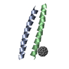
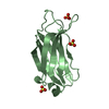
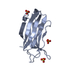
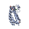
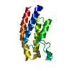
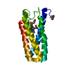
| Title | Crystal Structure of De novo Designed Fullerene organizing peptide | ||||||
 Components Components | Fullerene Organizing Protein (C60Sol-COP-3) | ||||||
 Keywords Keywords | DE NOVO PROTEIN / fullerene / complex / helical assembly | ||||||
| Function / homology | (C_{60}-I_{h})[5,6]fullerene Function and homology information Function and homology information | ||||||
| Biological species | synthetic construct (others) | ||||||
| Method |  X-RAY DIFFRACTION / SYNCHROTRON / MOLECULAR REPLACEMENT / molecular replacement / Resolution: 1.671 Å X-RAY DIFFRACTION / SYNCHROTRON / MOLECULAR REPLACEMENT / molecular replacement / Resolution: 1.671 Å | ||||||
 Authors Authors | Kim, K.-H. / Kim, Y.H. / Acharya, R. / Kim, N.H. / Paul, J. / Grigoryan, G. / DeGrado, W.F. | ||||||
| Funding support |  Korea, Republic Of, 1items Korea, Republic Of, 1items
| ||||||
 Citation Citation | Journal: Nat Commun / Year: 2016 Title: Protein-directed self-assembly of a fullerene crystal. Authors: Kim, K.-H. / Ko, D.-K. / Kim, Y.-T. / Kim, N.H. / Paul, J. / Zhang, S.-Q. / Murray, C.B. / Acharya, R. / DeGrado, W.F. / Kim, Y.H. / Grigoryan, G. | ||||||
| History |
|
- Structure visualization
| Structure viewer | Molecule: MolmilJmol/JSmol |
|---|
- Downloads & links
-Download
| PDBx/mmCIF format | 5et3.cif.gz | 35.6 KB | Display | PDBx/mmCIF format |
|---|---|---|---|---|
| PDB format | pdb5et3.ent.gz | 25.4 KB | Display | PDB format |
| PDBx/mmJSON format | 5et3.json.gz | Tree view | PDBx/mmJSON format | |
| Others |  Other downloads Other downloads |
-Validation report
| Arichive directory | https://data.pdbj.org/pub/pdb/validation_reports/et/5et3ftp://data.pdbj.org/pub/pdb/validation_reports/et/5et3 | HTTPS FTP |
|---|
-Related structure data
| Related structure data |  5hknC  5hkrC  3s0rS S: Starting model for refinement C: citing same article ( |
|---|---|
| Similar structure data |


-Links
 PDBj
PDBj

- Assembly
| Deposited unit | 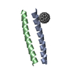
| ||||||||
|---|---|---|---|---|---|---|---|---|---|
| 1 | 
| ||||||||
| Unit cell |
|
-Components
| #1: Protein/peptide | Mass: 3188.565 Da / Num. of mol.: 2 / Source method: obtained synthetically / Source: (synth.) synthetic construct (others) #2: Chemical | ChemComp-60C / ( |   Mass: 720.642 Da / Num. of mol.: 1 / Source method: obtained synthetically / Formula: C60 Mass: 720.642 Da / Num. of mol.: 1 / Source method: obtained synthetically / Formula: C60#3: Water | ChemComp-HOH / |  Mass: 18.015 Da / Num. of mol.: 29 / Source method: isolated from a natural source / Formula: H2O Mass: 18.015 Da / Num. of mol.: 29 / Source method: isolated from a natural source / Formula: H2O |
|---|
-Experimental details
-Experiment
| Experiment | Method: X-RAY DIFFRACTION / Number of used crystals: 1 |
|---|
- Sample preparation
| Crystal | Density Matthews: 2.68 Å3/Da / Density % sol: 54.19 % |
|---|---|
| Crystal grow | Temperature: 295 K / Method: vapor diffusion, hanging drop / pH: 5.6 Details: The 2 uL drop consisted of a 1:1 v/v mixture of 8 mg/ml complex solution in 25 mM Tris pH 8.0 and reservoir solution consisting of 0.2 M Ammonium acetate, 0.1 M Sodium citrate tribasic ...Details: The 2 uL drop consisted of a 1:1 v/v mixture of 8 mg/ml complex solution in 25 mM Tris pH 8.0 and reservoir solution consisting of 0.2 M Ammonium acetate, 0.1 M Sodium citrate tribasic dihydrate pH 5.6, 30% w/v Polyethylene glycol 4,000 |
-Data collection
| Diffraction | Mean temperature: 100 K | ||||||||||||||||||||||||||||||||||||||||||||||||||||||||||||||||||
|---|---|---|---|---|---|---|---|---|---|---|---|---|---|---|---|---|---|---|---|---|---|---|---|---|---|---|---|---|---|---|---|---|---|---|---|---|---|---|---|---|---|---|---|---|---|---|---|---|---|---|---|---|---|---|---|---|---|---|---|---|---|---|---|---|---|---|---|
| Diffraction source | Source: SYNCHROTRON / Site: PAL/PLS / Beamline: 7A (6B, 6C1) / Wavelength: 1 Å | ||||||||||||||||||||||||||||||||||||||||||||||||||||||||||||||||||
| Detector | Type: ADSC QUANTUM 270 / Detector: CCD / Date: Oct 21, 2014 | ||||||||||||||||||||||||||||||||||||||||||||||||||||||||||||||||||
| Diffraction measurement | Details: 1.00 degrees, 1.0 sec, detector distance 200.00 mm / Method: \w scans | ||||||||||||||||||||||||||||||||||||||||||||||||||||||||||||||||||
| Radiation | Monochromator: double crystal monochromator / Protocol: SINGLE WAVELENGTH / Monochromatic (M) / Laue (L): M / Scattering type: x-ray | ||||||||||||||||||||||||||||||||||||||||||||||||||||||||||||||||||
| Radiation wavelength | Wavelength: 1 Å / Relative weight: 1 | ||||||||||||||||||||||||||||||||||||||||||||||||||||||||||||||||||
| Reflection | Av R equivalents: 0.066 / Number: 122822 | ||||||||||||||||||||||||||||||||||||||||||||||||||||||||||||||||||
| Reflection | Resolution: 1.67→50 Å / Num. obs: 7537 / % possible obs: 95.9 % / Redundancy: 16.3 % / Biso Wilson estimate: 11.04 Å2 / Rmerge(I) obs: 0.066 / Χ2: 0.947 / Net I/av σ(I): 30.99 / Net I/σ(I): 17.5 / Num. measured all: 122822 | ||||||||||||||||||||||||||||||||||||||||||||||||||||||||||||||||||
| Reflection shell | Diffraction-ID: 1 / Rejects: _
| ||||||||||||||||||||||||||||||||||||||||||||||||||||||||||||||||||
| Cell measurement | Reflection used: 122822 |
-Phasing
| Phasing | Method: molecular replacement |
|---|
- Processing
| Software |
| |||||||||||||||||||||||||||||||||||||||||||||||||||||||||||||||||||||||||||
|---|---|---|---|---|---|---|---|---|---|---|---|---|---|---|---|---|---|---|---|---|---|---|---|---|---|---|---|---|---|---|---|---|---|---|---|---|---|---|---|---|---|---|---|---|---|---|---|---|---|---|---|---|---|---|---|---|---|---|---|---|---|---|---|---|---|---|---|---|---|---|---|---|---|---|---|---|
| Refinement | Method to determine structure: MOLECULAR REPLACEMENT Starting model: 3S0R Resolution: 1.671→15.304 Å / SU ML: 0.16 / Cross valid method: THROUGHOUT / σ(F): 1.52 / Phase error: 24.14 / Stereochemistry target values: ML
| |||||||||||||||||||||||||||||||||||||||||||||||||||||||||||||||||||||||||||
| Solvent computation | Shrinkage radii: 0.9 Å / VDW probe radii: 1.11 Å / Solvent model: FLAT BULK SOLVENT MODEL | |||||||||||||||||||||||||||||||||||||||||||||||||||||||||||||||||||||||||||
| Displacement parameters | Biso max: 79.13 Å2 / Biso mean: 22.5022 Å2 / Biso min: 9.15 Å2 | |||||||||||||||||||||||||||||||||||||||||||||||||||||||||||||||||||||||||||
| Refinement step | Cycle: final / Resolution: 1.671→15.304 Å
| |||||||||||||||||||||||||||||||||||||||||||||||||||||||||||||||||||||||||||
| Refine LS restraints |
| |||||||||||||||||||||||||||||||||||||||||||||||||||||||||||||||||||||||||||
| LS refinement shell | Refine-ID: X-RAY DIFFRACTION / Total num. of bins used: 5
| |||||||||||||||||||||||||||||||||||||||||||||||||||||||||||||||||||||||||||
| Refinement TLS params. | Method: refined / Refine-ID: X-RAY DIFFRACTION
| |||||||||||||||||||||||||||||||||||||||||||||||||||||||||||||||||||||||||||
| Refinement TLS group |
|
