 Movie
Movie Controller


+ Open data

- Basic information
| Entry | Database: PDB / ID: 3s0r | ||||||
|---|---|---|---|---|---|---|---|
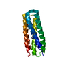
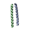
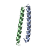
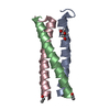
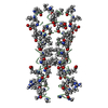
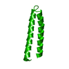
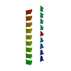
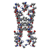
| Title | Crystal Structure of De novo Designed helical assembly protein | ||||||
 Components Components | De novo designed Helical Assembly | ||||||
 Keywords Keywords | DE NOVO PROTEIN / Interacts with Single Walled Nanotube (SWNT) | ||||||
| Biological species | artificial gene (others) | ||||||
| Method |  X-RAY DIFFRACTION / MOLECULAR REPLACEMENT / Resolution: 2.453 Å X-RAY DIFFRACTION / MOLECULAR REPLACEMENT / Resolution: 2.453 Å | ||||||
 Authors Authors | Acharya, R. / Grigoryan, G. / Kim, Y.H. / DeGrado, W.F. | ||||||
 Citation Citation | Journal: Science / Year: 2011 Title: Computational design of virus-like protein assemblies on carbon nanotube surfaces. Authors: Grigoryan, G. / Kim, Y.H. / Acharya, R. / Axelrod, K. / Jain, R.M. / Willis, L. / Drndic, M. / Kikkawa, J.M. / DeGrado, W.F. | ||||||
| History |
|
- Structure visualization
| Structure viewer | Molecule:  MolmilJmol/JSmol MolmilJmol/JSmol |
|---|
- Downloads & links
-Download
| PDBx/mmCIF format | 3s0r.cif.gz | 19.2 KB | Display | PDBx/mmCIF format |
|---|---|---|---|---|
| PDB format | pdb3s0r.ent.gz | 13.6 KB | Display | PDB format |
| PDBx/mmJSON format | 3s0r.json.gz | Tree view | PDBx/mmJSON format | |
| Others |  Other downloads Other downloads |
-Validation report
| Arichive directory | https://data.pdbj.org/pub/pdb/validation_reports/s0/3s0rftp://data.pdbj.org/pub/pdb/validation_reports/s0/3s0r | HTTPS FTP |
|---|
-Related structure data
| Similar structure data |
|---|


-Links
 PDBj
PDBj
- Assembly
| Deposited unit | 
| ||||||||
|---|---|---|---|---|---|---|---|---|---|
| 1 | 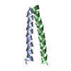
| ||||||||
| Unit cell |
|
-Components
| #1: Protein/peptide | Mass: 3188.565 Da / Num. of mol.: 2 / Source method: obtained synthetically / Source: (synth.) artificial gene (others) #2: Water | ChemComp-HOH / |  Mass: 18.015 Da / Num. of mol.: 18 / Source method: isolated from a natural source / Formula: H2O Mass: 18.015 Da / Num. of mol.: 18 / Source method: isolated from a natural source / Formula: H2O |
|---|
-Experimental details
-Experiment
| Experiment | Method: X-RAY DIFFRACTION / Number of used crystals: 1 |
|---|
- Sample preparation
| Crystal | Density Matthews: 3.79 Å3/Da / Density % sol: 67.55 % |
|---|---|
| Crystal grow | Temperature: 291 K / Method: vapor diffusion, hanging drop / pH: 7.5 Details: The 2 uL drop consisted of a 1:1 v/v mixture of 5 mg/mL protein solution (in 20 mM sodium phosphate, pH 7.5, 100 mM NaCl) and reservoir solution (75 mM HEPES sodium, pH 7.5, 0.6 M sodium ...Details: The 2 uL drop consisted of a 1:1 v/v mixture of 5 mg/mL protein solution (in 20 mM sodium phosphate, pH 7.5, 100 mM NaCl) and reservoir solution (75 mM HEPES sodium, pH 7.5, 0.6 M sodium phosphate monobasic monohydrate, 0.6 M potassium phosphate monobasic, 25% v/v glycerol) , VAPOR DIFFUSION, HANGING DROP, temperature 291K |
-Data collection
| Diffraction | Mean temperature: 100 K |
|---|---|
| Diffraction source | Source: ROTATING ANODE / Type: RIGAKU / Wavelength: 1.5418 |
| Detector | Type: MAR scanner 300 mm plate / Detector: IMAGE PLATE / Date: May 21, 2010 / Details: mirrors |
| Radiation | Protocol: SINGLE WAVELENGTH / Monochromatic (M) / Laue (L): M / Scattering type: x-ray |
| Radiation wavelength | Wavelength: 1.5418 Å / Relative weight: 1 |
| Reflection twin | Operator: h,-k,-l / Fraction: 0.499 |
| Reflection | Resolution: 2.44→18.888 Å / Num. all: 3501 / Num. obs: 3479 / % possible obs: 89.5 % / Observed criterion σ(I): 3 / Redundancy: 7.4 % / Biso Wilson estimate: 38.2 Å2 / Rmerge(I) obs: 0.067 / Net I/σ(I): 20.5 |
| Reflection shell | Resolution: 2.44→2.57 Å / Redundancy: 7.1 % / Rmerge(I) obs: 0.168 / Mean I/σ(I) obs: 10.8 / Num. unique all: 434 / % possible all: 81.8 |
- Processing
| Software |
| |||||||||||||||||||||||||
|---|---|---|---|---|---|---|---|---|---|---|---|---|---|---|---|---|---|---|---|---|---|---|---|---|---|---|
| Refinement | Method to determine structure: MOLECULAR REPLACEMENT Starting model: 30-residue polyalanine alpha helix Resolution: 2.453→18.888 Å / Cross valid method: THROUGHOUT / σ(F): 2.07 / Phase error: 30.11 / Stereochemistry target values: TWIN_LSQ_F
| |||||||||||||||||||||||||
| Solvent computation | Shrinkage radii: 0.9 Å / VDW probe radii: 1.11 Å / Solvent model: FLAT BULK SOLVENT MODEL / Bsol: 78.95 Å2 / ksol: 0.5 e/Å3 | |||||||||||||||||||||||||
| Displacement parameters | Biso mean: 28.2 Å2
| |||||||||||||||||||||||||
| Refinement step | Cycle: LAST / Resolution: 2.453→18.888 Å
| |||||||||||||||||||||||||
| Refine LS restraints |
| |||||||||||||||||||||||||
| LS refinement shell | Refine-ID: X-RAY DIFFRACTION / Total num. of bins used: 2 / % reflection obs: 91 %
|
