 Movie
Movie Controller

[English] 日本語
 Yorodumi
- PDB-5elz: Staphylococcus aureus Type II pantothenate kinase in complex with... -
+ Open data

- Basic information
| Entry | Database: PDB / ID: 5elz | ||||||
|---|---|---|---|---|---|---|---|
| Title | Staphylococcus aureus Type II pantothenate kinase in complex with a pantothenate analog | ||||||
 Components Components | Type II pantothenate kinase | ||||||
 Keywords Keywords | Transferase/Transferase Inhibitor / Pantothenate kinase / Structural Genomics / Structural Genomics Consortium / SGC / Transferase-Transferase Inhibitor complex | ||||||
| Function / homology |  Function and homology information Function and homology informationpantothenate kinase / pantothenate kinase activity / coenzyme A biosynthetic process / ATP binding / cytosol Similarity search - Function | ||||||
| Biological species |   Staphylococcus aureus (bacteria) Staphylococcus aureus (bacteria) | ||||||
| Method |  X-RAY DIFFRACTION / MOLECULAR REPLACEMENT / Resolution: 1.8 Å X-RAY DIFFRACTION / MOLECULAR REPLACEMENT / Resolution: 1.8 Å | ||||||
 Authors Authors | Mottaghi, K. / Hughes, S.J. / Tempel, W. / Hong, B. / Park, H. / Structural Genomics Consortium (SGC) | ||||||
 Citation Citation | Journal: Acs Infect Dis. / Year: 2016 Title: Discovery of Potent Pantothenamide Inhibitors of Staphylococcus aureus Pantothenate Kinase through a Minimal SAR Study: Inhibition Is Due to Trapping of the Product. Authors: Hughes, S.J. / Barnard, L. / Mottaghi, K. / Tempel, W. / Antoshchenko, T. / Hong, B.S. / Allali-Hassani, A. / Smil, D. / Vedadi, M. / Strauss, E. / Park, H.W. | ||||||
| History |
|
- Structure visualization
| Structure viewer | Molecule: MolmilJmol/JSmol |
|---|
- Downloads & links
-Download
| PDBx/mmCIF format | 5elz.cif.gz | 131 KB | Display | PDBx/mmCIF format |
|---|---|---|---|---|
| PDB format | pdb5elz.ent.gz | 99.3 KB | Display | PDB format |
| PDBx/mmJSON format | 5elz.json.gz | Tree view | PDBx/mmJSON format | |
| Others |  Other downloads Other downloads |
-Validation report
| Arichive directory | https://data.pdbj.org/pub/pdb/validation_reports/el/5elzftp://data.pdbj.org/pub/pdb/validation_reports/el/5elz | HTTPS FTP |
|---|
-Related structure data
| Related structure data |  4m7xSC  4m7yC  5jicC S: Starting model for refinement C: citing same article ( |
|---|---|
| Similar structure data |
-Links
 PDBj
PDBj- Assembly
| Deposited unit | 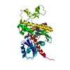
| ||||||||
|---|---|---|---|---|---|---|---|---|---|
| 1 | 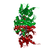
| ||||||||
| Unit cell |
| ||||||||
| Components on special symmetry positions |
|
-Components
-Protein , 1 types, 1 molecules A
| #1: Protein | Mass: 29955.818 Da / Num. of mol.: 1 Source method: isolated from a genetically manipulated source Source: (gene. exp.) Staphylococcus aureus (bacteria) / Strain: MW2 / Gene: coaW, MW2054 / Production host: References: UniProt: Q8NVG0, UniProt: Q2FWC7*PLUS, pantothenate kinase |
|---|
-Non-polymers , 5 types, 280 molecules 


| #2: Chemical | ChemComp-ADP /  Mass: 427.201 Da / Num. of mol.: 1 / Source method: obtained synthetically / Formula: C10H15N5O10P2 / Comment: ADP, energy-carrying molecule*YM Mass: 427.201 Da / Num. of mol.: 1 / Source method: obtained synthetically / Formula: C10H15N5O10P2 / Comment: ADP, energy-carrying molecule*YM | ||
|---|---|---|---|
| #3: Chemical | ChemComp-MG /  Mass: 24.305 Da / Num. of mol.: 1 / Source method: obtained synthetically / Formula: Mg Mass: 24.305 Da / Num. of mol.: 1 / Source method: obtained synthetically / Formula: Mg | ||
| #4: Chemical | ChemComp-3V9 /  Mass: 432.362 Da / Num. of mol.: 1 / Source method: obtained synthetically / Formula: C17H25N2O9P Mass: 432.362 Da / Num. of mol.: 1 / Source method: obtained synthetically / Formula: C17H25N2O9P | ||
| #5: Chemical |  Num. of mol.: 2 / Source method: obtained synthetically Num. of mol.: 2 / Source method: obtained synthetically#6: Water | ChemComp-HOH / | Mass: 18.015 Da / Num. of mol.: 275 / Source method: isolated from a natural source / Formula: H2O |
-Experimental details
-Experiment
| Experiment | Method: X-RAY DIFFRACTION / Number of used crystals: 1 |
|---|
- Sample preparation
| Crystal | Density Matthews: 2.4 Å3/Da / Density % sol: 48.79 % |
|---|---|
| Crystal grow | Temperature: 291 K / Method: vapor diffusion / pH: 8.5 Details: 0.5 ul protein + 0.5 uL buffer that contains 25% PEG3350, 0.2 M MgCl2, 0.1 M Tris HCl, pH 8.5 |
-Data collection
| Diffraction | Mean temperature: 100 K |
|---|---|
| Diffraction source | Source: ROTATING ANODE / Type: RIGAKU / Wavelength: 1.5418 Å |
| Detector | Type: RIGAKU SATURN A200 / Detector: CCD / Date: Mar 19, 2012 |
| Radiation | Protocol: SINGLE WAVELENGTH / Monochromatic (M) / Laue (L): M / Scattering type: x-ray |
| Radiation wavelength | Wavelength: 1.5418 Å / Relative weight: 1 |
| Reflection | Resolution: 1.8→28.48 Å / Num. obs: 27062 / % possible obs: 99.9 % / Redundancy: 7 % / Rmerge(I) obs: 0.058 / Net I/σ(I): 23.1 |
| Reflection shell | Resolution: 1.8→1.9 Å / Redundancy: 6.8 % / Rmerge(I) obs: 0.498 / Mean I/σ(I) obs: 4 / % possible all: 99.2 |
- Processing
| Software |
| |||||||||||||||||||||||||||||||||||||||||||||||||||||||||||||||||||||||||||
|---|---|---|---|---|---|---|---|---|---|---|---|---|---|---|---|---|---|---|---|---|---|---|---|---|---|---|---|---|---|---|---|---|---|---|---|---|---|---|---|---|---|---|---|---|---|---|---|---|---|---|---|---|---|---|---|---|---|---|---|---|---|---|---|---|---|---|---|---|---|---|---|---|---|---|---|---|
| Refinement | Method to determine structure: MOLECULAR REPLACEMENT Starting model: PDB entry 4M7X Resolution: 1.8→28 Å / Cor.coef. Fo:Fc: 0.961 / Cor.coef. Fo:Fc free: 0.94 / WRfactor Rfree: 0.1936 / WRfactor Rwork: 0.1557 / FOM work R set: 0.8873 / SU B: 4.476 / SU ML: 0.08 / SU R Cruickshank DPI: 0.1225 / SU Rfree: 0.118 / Cross valid method: THROUGHOUT / σ(F): 0 / ESU R: 0.122 / ESU R Free: 0.118 / Stereochemistry target values: MAXIMUM LIKELIHOOD / Details: HYDROGENS HAVE BEEN ADDED IN THE RIDING POSITIONS
| |||||||||||||||||||||||||||||||||||||||||||||||||||||||||||||||||||||||||||
| Solvent computation | Ion probe radii: 0.8 Å / Shrinkage radii: 0.8 Å / VDW probe radii: 1.2 Å / Solvent model: MASK | |||||||||||||||||||||||||||||||||||||||||||||||||||||||||||||||||||||||||||
| Displacement parameters | Biso max: 85.83 Å2 / Biso mean: 24.876 Å2 / Biso min: 9.84 Å2
| |||||||||||||||||||||||||||||||||||||||||||||||||||||||||||||||||||||||||||
| Refinement step | Cycle: final / Resolution: 1.8→28 Å
| |||||||||||||||||||||||||||||||||||||||||||||||||||||||||||||||||||||||||||
| Refine LS restraints |
| |||||||||||||||||||||||||||||||||||||||||||||||||||||||||||||||||||||||||||
| LS refinement shell | Resolution: 1.8→1.848 Å / Total num. of bins used: 20
| |||||||||||||||||||||||||||||||||||||||||||||||||||||||||||||||||||||||||||
| Refinement TLS params. | Method: refined / Origin x: 17.2881 Å / Origin y: 14.3542 Å / Origin z: -3.5316 Å
|
