 Movie
Movie Controller

[English] 日本語
 Yorodumi
- PDB-5edj: Crystal structure of the Neisseria meningitidis iron-regulated ou... -
+ Open data

- Basic information
| Entry | Database: PDB / ID: 5edj | ||||||
|---|---|---|---|---|---|---|---|
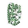
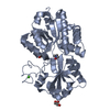
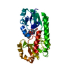
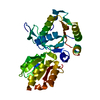
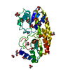
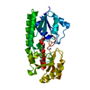
| Title | Crystal structure of the Neisseria meningitidis iron-regulated outer membrane lipoprotein FrpD | ||||||
 Components Components | FrpC operon protein | ||||||
 Keywords Keywords | UNKNOWN FUNCTION / Iron-regulated protein FrpD / FrpC-binding protein / novel fold / membrane protein | ||||||
| Function / homology | RTX iron-regulated FrpC / RTX iron-regulated protein FrpC / Prokaryotic membrane lipoprotein lipid attachment site profile. / RTX iron-regulated protein (frpC) / FrpC operon protein Function and homology information Function and homology information | ||||||
| Biological species |  Neisseria meningitidis MC58 (bacteria) Neisseria meningitidis MC58 (bacteria) | ||||||
| Method |  X-RAY DIFFRACTION / SYNCHROTRON / MOLECULAR REPLACEMENT / Resolution: 2.3 Å X-RAY DIFFRACTION / SYNCHROTRON / MOLECULAR REPLACEMENT / Resolution: 2.3 Å | ||||||
 Authors Authors | Sviridova, E. / Bumba, L. / Rezacova, P. / Sebo, P. / Kuta Smatanova, I. | ||||||
| Funding support |  Czech Republic, 1items Czech Republic, 1items
| ||||||
 Citation Citation | Journal: Sci Rep / Year: 2017 Title: Structural basis of the interaction between the putative adhesion-involved and iron-regulated FrpD and FrpC proteins of Neisseria meningitidis. Authors: Sviridova, E. / Rezacova, P. / Bondar, A. / Veverka, V. / Novak, P. / Schenk, G. / Svergun, D.I. / Kuta Smatanova, I. / Bumba, L. #1: Journal: Acta Crystallographica Section F / Year: 2010 Title: Crystallization and preliminary crystallographic characterization of the iron-regulated outer membrane lipoprotein FrpD from Neisseria meningitidis Authors: Sviridova, E. / Bumba, L. / Rezacova, P. / Prochazkova, K. / Kavan, D. / Bezoushka, K. / Kuty, M. / Sebo, P. / Kuta Smatanova, I. | ||||||
| History |
|
- Structure visualization
| Structure viewer | Molecule: MolmilJmol/JSmol |
|---|
- Downloads & links
-Download
| PDBx/mmCIF format | 5edj.cif.gz | 113.9 KB | Display | PDBx/mmCIF format |
|---|---|---|---|---|
| PDB format | pdb5edj.ent.gz | 87.6 KB | Display | PDB format |
| PDBx/mmJSON format | 5edj.json.gz | Tree view | PDBx/mmJSON format | |
| Others |  Other downloads Other downloads |
-Validation report
| Arichive directory | https://data.pdbj.org/pub/pdb/validation_reports/ed/5edjftp://data.pdbj.org/pub/pdb/validation_reports/ed/5edj | HTTPS FTP |
|---|
-Related structure data
| Related structure data | 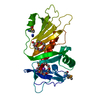 5edfSC S: Starting model for refinement C: citing same article ( |
|---|---|
| Similar structure data |


-Links
 PDBj
PDBj


- Assembly
| Deposited unit | 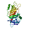
| ||||||||
|---|---|---|---|---|---|---|---|---|---|
| 1 |
| ||||||||
| Unit cell |
|
-Components
| #1: Protein | Mass: 27027.986 Da / Num. of mol.: 1 / Fragment: UNP residues 43-271 Source method: isolated from a genetically manipulated source Details: Fragment FrpD43-271, where: 1. the sequence position present in the structure is 44-267; 2. LE sequence at the C-terminus is a cloning artifact Source: (gene. exp.) Neisseria meningitidis MC58 (bacteria) / Gene: LA50_03295 / Plasmid: pET28b / Production host: |
|---|---|
| #2: Water | ChemComp-HOH /  Mass: 18.015 Da / Num. of mol.: 130 / Source method: isolated from a natural source / Formula: H2O Mass: 18.015 Da / Num. of mol.: 130 / Source method: isolated from a natural source / Formula: H2O |
| Has protein modification | Y |
-Experimental details
-Experiment
| Experiment | Method: X-RAY DIFFRACTION |
|---|
- Sample preparation
| Crystal | Density Matthews: 2.85 Å3/Da / Density % sol: 56.8 % |
|---|---|
| Crystal grow | Temperature: 293 K / Method: vapor diffusion, sitting drop / pH: 8.5 / Details: 0.1 M Tris-HCl, 2 M ammonium sulfate |
-Data collection
| Diffraction | Mean temperature: 100 K |
|---|---|
| Diffraction source | Source: SYNCHROTRON / Site: BESSY  / Beamline: 14.2 / Wavelength: 0.918 Å / Beamline: 14.2 / Wavelength: 0.918 Å |
| Detector | Type: RAYONIX MX-225 / Detector: CCD / Date: Oct 23, 2008 |
| Radiation | Protocol: SINGLE WAVELENGTH / Monochromatic (M) / Laue (L): M / Scattering type: x-ray |
| Radiation wavelength | Wavelength: 0.918 Å / Relative weight: 1 |
| Reflection | Resolution: 2.3→50 Å / Num. all: 13418 / Num. obs: 13324 / % possible obs: 99.3 % / Observed criterion σ(F): 0 / Observed criterion σ(I): -3 / Redundancy: 6.9 % / Biso Wilson estimate: 33.2 Å2 / Rmerge(I) obs: 0.071 / Net I/σ(I): 22.9 |
| Reflection shell | Resolution: 2.3→2.38 Å / Redundancy: 4.9 % / % possible all: 95.1 |
- Processing
| Software |
| ||||||||||||||||||||||||||||||||||||||||||||||||||||||||||||||||||||||||||||||||||||||||||||||||||||||||||||||||||||||||||||||||||||||||||||||||||||||||||||||||||||||||||||||||||||||
|---|---|---|---|---|---|---|---|---|---|---|---|---|---|---|---|---|---|---|---|---|---|---|---|---|---|---|---|---|---|---|---|---|---|---|---|---|---|---|---|---|---|---|---|---|---|---|---|---|---|---|---|---|---|---|---|---|---|---|---|---|---|---|---|---|---|---|---|---|---|---|---|---|---|---|---|---|---|---|---|---|---|---|---|---|---|---|---|---|---|---|---|---|---|---|---|---|---|---|---|---|---|---|---|---|---|---|---|---|---|---|---|---|---|---|---|---|---|---|---|---|---|---|---|---|---|---|---|---|---|---|---|---|---|---|---|---|---|---|---|---|---|---|---|---|---|---|---|---|---|---|---|---|---|---|---|---|---|---|---|---|---|---|---|---|---|---|---|---|---|---|---|---|---|---|---|---|---|---|---|---|---|---|---|
| Refinement | Method to determine structure: MOLECULAR REPLACEMENT Starting model: 5EDF Resolution: 2.3→30.65 Å / Cor.coef. Fo:Fc: 0.946 / Cor.coef. Fo:Fc free: 0.913 / SU B: 15.477 / SU ML: 0.167 / Cross valid method: THROUGHOUT / ESU R: 0.278 / ESU R Free: 0.231 / Stereochemistry target values: MAXIMUM LIKELIHOOD / Details: HYDROGENS HAVE BEEN ADDED IN THE RIDING POSITIONS
| ||||||||||||||||||||||||||||||||||||||||||||||||||||||||||||||||||||||||||||||||||||||||||||||||||||||||||||||||||||||||||||||||||||||||||||||||||||||||||||||||||||||||||||||||||||||
| Solvent computation | Ion probe radii: 0.8 Å / Shrinkage radii: 0.8 Å / VDW probe radii: 1.4 Å / Solvent model: BABINET MODEL WITH MASK | ||||||||||||||||||||||||||||||||||||||||||||||||||||||||||||||||||||||||||||||||||||||||||||||||||||||||||||||||||||||||||||||||||||||||||||||||||||||||||||||||||||||||||||||||||||||
| Displacement parameters | Biso mean: 41.071 Å2
| ||||||||||||||||||||||||||||||||||||||||||||||||||||||||||||||||||||||||||||||||||||||||||||||||||||||||||||||||||||||||||||||||||||||||||||||||||||||||||||||||||||||||||||||||||||||
| Refinement step | Cycle: 1 / Resolution: 2.3→30.65 Å
| ||||||||||||||||||||||||||||||||||||||||||||||||||||||||||||||||||||||||||||||||||||||||||||||||||||||||||||||||||||||||||||||||||||||||||||||||||||||||||||||||||||||||||||||||||||||
| Refine LS restraints |
|