 Movie
Movie Controller

[English] 日本語
 Yorodumi
- PDB-5e9h: Structural insights of isocitrate lyases from Fusarium graminearum -
+ Open data

- Basic information
| Entry | Database: PDB / ID: 5e9h | ||||||
|---|---|---|---|---|---|---|---|
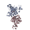
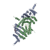
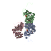
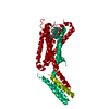
| Title | Structural insights of isocitrate lyases from Fusarium graminearum | ||||||
 Components Components | Isocitrate lyase | ||||||
 Keywords Keywords | LYASE / TIM BETA/ALPHA-BARREL / LYASE ACTIVITY | ||||||
| Function / homology |  Function and homology information Function and homology informationglyoxysome / methylisocitrate lyase / isocitrate lyase / isocitrate lyase activity / methylisocitrate lyase activity / glyoxylate cycle / tricarboxylic acid cycle / metal ion binding Similarity search - Function | ||||||
| Biological species |  Gibberella zeae PH-1 (fungus) Gibberella zeae PH-1 (fungus) | ||||||
| Method |  X-RAY DIFFRACTION / SYNCHROTRON / MOLECULAR REPLACEMENT / Resolution: 2.3 Å X-RAY DIFFRACTION / SYNCHROTRON / MOLECULAR REPLACEMENT / Resolution: 2.3 Å | ||||||
 Authors Authors | Park, Y. / Cho, Y. / Lee, Y.-H. / Lee, Y.-W. / Rhee, S. | ||||||
 Citation Citation | Journal: J.Struct.Biol. / Year: 2016 Title: Crystal structure and functional analysis of isocitrate lyases from Magnaporthe oryzae and Fusarium graminearum Authors: Park, Y. / Cho, Y. / Lee, Y.-H. / Lee, Y.-W. / Rhee, S. | ||||||
| History |
|
- Structure visualization
| Structure viewer | Molecule: MolmilJmol/JSmol |
|---|
- Downloads & links
-Download
| PDBx/mmCIF format | 5e9h.cif.gz | 223.9 KB | Display | PDBx/mmCIF format |
|---|---|---|---|---|
| PDB format | pdb5e9h.ent.gz | 177.4 KB | Display | PDB format |
| PDBx/mmJSON format | 5e9h.json.gz | Tree view | PDBx/mmJSON format | |
| Others |  Other downloads Other downloads |
-Validation report
| Arichive directory | https://data.pdbj.org/pub/pdb/validation_reports/e9/5e9hftp://data.pdbj.org/pub/pdb/validation_reports/e9/5e9h | HTTPS FTP |
|---|
-Related structure data
| Related structure data |  5e9fC  5e9gC 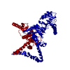 1dquS C: citing same article ( S: Starting model for refinement |
|---|---|
| Similar structure data |


-Links
 PDBj
PDBj
- Assembly
| Deposited unit | 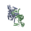
| ||||||||||||||||||
|---|---|---|---|---|---|---|---|---|---|---|---|---|---|---|---|---|---|---|---|
| 1 |
| ||||||||||||||||||
| Unit cell |
| ||||||||||||||||||
| Components on special symmetry positions |
|
-Components
| #1: Protein | Mass: 61971.938 Da / Num. of mol.: 2 Source method: isolated from a genetically manipulated source Source: (gene. exp.) Gibberella zeae PH-1 (fungus) / Strain: PH-1 / Gene: ICL1, FGSG_09896 / Production host:  References: UniProt: Q4HYR2, isocitrate lyase, methylisocitrate lyase #2: Chemical | ChemComp-MN / |   Mass: 54.938 Da / Num. of mol.: 1 / Source method: obtained synthetically / Formula: Mn Mass: 54.938 Da / Num. of mol.: 1 / Source method: obtained synthetically / Formula: Mn#3: Chemical | ChemComp-MLI / |   Mass: 102.046 Da / Num. of mol.: 1 / Source method: obtained synthetically / Formula: C3H2O4 Mass: 102.046 Da / Num. of mol.: 1 / Source method: obtained synthetically / Formula: C3H2O4#4: Water | ChemComp-HOH / |  Mass: 18.015 Da / Num. of mol.: 420 / Source method: isolated from a natural source / Formula: H2O Mass: 18.015 Da / Num. of mol.: 420 / Source method: isolated from a natural source / Formula: H2O |
|---|
-Experimental details
-Experiment
| Experiment | Method: X-RAY DIFFRACTION / Number of used crystals: 1 |
|---|
- Sample preparation
| Crystal | Density Matthews: 2.94 Å3/Da / Density % sol: 58.22 % |
|---|---|
| Crystal grow | Temperature: 295 K / Method: vapor diffusion, sitting drop / pH: 8 Details: 2.4M SODIUM MALONATE, PH 8.0, VAPOR DIFFUSION, SITTING DROP, TEMPERATURE 295.0K |
-Data collection
| Diffraction | Mean temperature: 100 K |
|---|---|
| Diffraction source | Source: SYNCHROTRON / Site: PAL/PLS  / Beamline: 7A (6B, 6C1) / Wavelength: 0.979 Å / Beamline: 7A (6B, 6C1) / Wavelength: 0.979 Å |
| Detector | Type: ADSC QUANTUM 270 / Detector: CCD / Date: Oct 1, 2012 |
| Radiation | Protocol: SINGLE WAVELENGTH / Monochromatic (M) / Laue (L): M / Scattering type: x-ray |
| Radiation wavelength | Wavelength: 0.979 Å / Relative weight: 1 |
| Reflection | Resolution: 2.3→50 Å / Num. obs: 64136 / % possible obs: 99.95 % / Redundancy: 21.2 % / Net I/σ(I): 19.87 |
| Reflection shell | Resolution: 2.3→2.38 Å |
- Processing
| Software |
| |||||||||||||||||||||||||||||||||||||||||||||||||||||||||||||||||||||||||||||||||||||||||||||||||||||||||
|---|---|---|---|---|---|---|---|---|---|---|---|---|---|---|---|---|---|---|---|---|---|---|---|---|---|---|---|---|---|---|---|---|---|---|---|---|---|---|---|---|---|---|---|---|---|---|---|---|---|---|---|---|---|---|---|---|---|---|---|---|---|---|---|---|---|---|---|---|---|---|---|---|---|---|---|---|---|---|---|---|---|---|---|---|---|---|---|---|---|---|---|---|---|---|---|---|---|---|---|---|---|---|---|---|---|---|
| Refinement | Method to determine structure: MOLECULAR REPLACEMENT Starting model: 1DQU Resolution: 2.3→32.4 Å / SU ML: 0.2 / Cross valid method: FREE R-VALUE / σ(F): 1.34 / Phase error: 24.23 / Stereochemistry target values: ML
| |||||||||||||||||||||||||||||||||||||||||||||||||||||||||||||||||||||||||||||||||||||||||||||||||||||||||
| Solvent computation | Shrinkage radii: 0.9 Å / VDW probe radii: 1.11 Å / Solvent model: FLAT BULK SOLVENT MODEL | |||||||||||||||||||||||||||||||||||||||||||||||||||||||||||||||||||||||||||||||||||||||||||||||||||||||||
| Refinement step | Cycle: LAST / Resolution: 2.3→32.4 Å
| |||||||||||||||||||||||||||||||||||||||||||||||||||||||||||||||||||||||||||||||||||||||||||||||||||||||||
| Refine LS restraints |
| |||||||||||||||||||||||||||||||||||||||||||||||||||||||||||||||||||||||||||||||||||||||||||||||||||||||||
| LS refinement shell |
|
