 Movie
Movie Controller

[English] 日本語
 Yorodumi
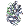
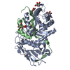
- PDB-5e9b: Crystal structure of human heparanase in complex with HepMer M09S05a -
+ Open data

- Basic information
| Entry | Database: PDB / ID: 5e9b | ||||||||||||
|---|---|---|---|---|---|---|---|---|---|---|---|---|---|
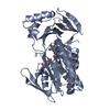
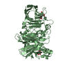
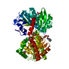
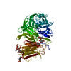
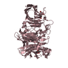
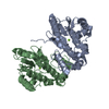
| Title | Crystal structure of human heparanase in complex with HepMer M09S05a | ||||||||||||
 Components Components | (Heparanase) x 2 | ||||||||||||
 Keywords Keywords | HYDROLASE / glycoside hydrolase / ligand 3 / protein / sugar | ||||||||||||
| Function / homology |  Function and homology information Function and homology informationheparanase / heparanase activity / regulation of hair follicle development / heparin proteoglycan metabolic process / heparan sulfate proteoglycan catabolic process / beta-glucuronidase activity / proteoglycan metabolic process / positive regulation of hair follicle development / syndecan binding / vascular wound healing ...heparanase / heparanase activity / regulation of hair follicle development / heparin proteoglycan metabolic process / heparan sulfate proteoglycan catabolic process / beta-glucuronidase activity / proteoglycan metabolic process / positive regulation of hair follicle development / syndecan binding / vascular wound healing / protein transmembrane transport / HS-GAG degradation / angiogenesis involved in wound healing / establishment of endothelial barrier / positive regulation of osteoblast proliferation / positive regulation of blood coagulation / positive regulation of vascular endothelial growth factor production / lysosomal lumen / cell-matrix adhesion / specific granule lumen / extracellular matrix / positive regulation of phosphatidylinositol 3-kinase/protein kinase B signal transduction / lysosome / membrane raft / lysosomal membrane / response to antibiotic / Neutrophil degranulation / : / extracellular region / nucleoplasm / nucleus / plasma membrane Similarity search - Function | ||||||||||||
| Biological species |  Homo sapiens (human) Homo sapiens (human) | ||||||||||||
| Method |  X-RAY DIFFRACTION / SYNCHROTRON / MOLECULAR REPLACEMENT / Resolution: 1.88 Å X-RAY DIFFRACTION / SYNCHROTRON / MOLECULAR REPLACEMENT / Resolution: 1.88 Å | ||||||||||||
 Authors Authors | Wu, L. / Davies, G.J. | ||||||||||||
 Citation Citation | Journal: Nat.Struct.Mol.Biol. / Year: 2015 Title: Structural characterization of human heparanase reveals insights into substrate recognition. Authors: Wu, L. / Viola, C.M. / Brzozowski, A.M. / Davies, G.J. | ||||||||||||
| History |
|
- Structure visualization
| Structure viewer | Molecule: MolmilJmol/JSmol |
|---|
- Downloads & links
-Download
| PDBx/mmCIF format | 5e9b.cif.gz | 205.7 KB | Display | PDBx/mmCIF format |
|---|---|---|---|---|
| PDB format | pdb5e9b.ent.gz | 158.5 KB | Display | PDB format |
| PDBx/mmJSON format | 5e9b.json.gz | Tree view | PDBx/mmJSON format | |
| Others |  Other downloads Other downloads |
-Validation report
| Arichive directory | https://data.pdbj.org/pub/pdb/validation_reports/e9/5e9bftp://data.pdbj.org/pub/pdb/validation_reports/e9/5e9b | HTTPS FTP |
|---|
-Related structure data
| Related structure data |  5e8mSC  5e97C 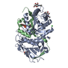 5e98C 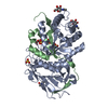 5e9cC S: Starting model for refinement C: citing same article ( |
|---|---|
| Similar structure data |


-Links
 PDBj
PDBj- Assembly
| Deposited unit | 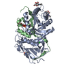
| ||||||||
|---|---|---|---|---|---|---|---|---|---|
| 1 |
| ||||||||
| Unit cell |
|
-Components
-Protein , 2 types, 2 molecules AB
| #1: Protein | Mass: 43733.324 Da / Num. of mol.: 1 / Fragment: residues 158-543 Source method: isolated from a genetically manipulated source Source: (gene. exp.) Homo sapiens (human) / Gene: HPSE, HEP, HPA, HPA1, HPR1, HPSE1, HSE1 / Production host:  Trichoplusia ni (cabbage looper) / References: UniProt: Q9Y251, heparanase Trichoplusia ni (cabbage looper) / References: UniProt: Q9Y251, heparanase |
|---|---|
| #2: Protein | Mass: 8542.769 Da / Num. of mol.: 1 / Fragment: residues 36-109 Source method: isolated from a genetically manipulated source Source: (gene. exp.) Homo sapiens (human) / Gene: HPSE, HEP, HPA, HPA1, HPR1, HPSE1, HSE1 / Production host: Trichoplusia ni (cabbage looper) / References: UniProt: Q9Y251, heparanase |
-Sugars , 2 types, 4 molecules 
| #3: Polysaccharide | 2-deoxy-2-(sulfoamino)-alpha-D-glucopyranose-(1-4)-beta-D-glucopyranuronic acid-(1-4)-2-deoxy-6-O- ...2-deoxy-2-(sulfoamino)-alpha-D-glucopyranose-(1-4)-beta-D-glucopyranuronic acid-(1-4)-2-deoxy-6-O-sulfo-2-(sulfoamino)-alpha-D-glucopyranose-(1-4)-beta-D-glucopyranuronic acid Source method: isolated from a genetically manipulated source |
|---|---|
| #4: Sugar |  Type: D-saccharide, beta linking / Mass: 221.208 Da / Num. of mol.: 3 Type: D-saccharide, beta linking / Mass: 221.208 Da / Num. of mol.: 3Source method: isolated from a genetically manipulated source Formula: C8H15NO6 |
-Non-polymers , 2 types, 202 molecules 

| #5: Chemical | ChemComp-CL /  Mass: 35.453 Da / Num. of mol.: 1 / Source method: obtained synthetically / Formula: Cl Mass: 35.453 Da / Num. of mol.: 1 / Source method: obtained synthetically / Formula: Cl |
|---|---|
| #6: Water | ChemComp-HOH / Mass: 18.015 Da / Num. of mol.: 201 / Source method: isolated from a natural source / Formula: H2O |
-Details
| Has ligand of interest | Y |
|---|---|
| Has protein modification | Y |
-Experimental details
-Experiment
| Experiment | Method: X-RAY DIFFRACTION |
|---|
- Sample preparation
| Crystal | Density Matthews: 2.32 Å3/Da / Density % sol: 47 % |
|---|---|
| Crystal grow | Temperature: 293 K / Method: vapor diffusion, sitting drop / pH: 5.5 / Details: 0.1 M MES [5.5] 0.1 M MgCl2 17% PEG3350 |
-Data collection
| Diffraction | Mean temperature: 100 K / Serial crystal experiment: N |
|---|---|
| Diffraction source | Source: SYNCHROTRON / Site: Diamond  / Beamline: I03 / Wavelength: 0.85 Å / Beamline: I03 / Wavelength: 0.85 Å |
| Detector | Type: DECTRIS PILATUS 6M / Detector: PIXEL / Date: Jul 4, 2015 |
| Radiation | Protocol: SINGLE WAVELENGTH / Monochromatic (M) / Laue (L): M / Scattering type: x-ray |
| Radiation wavelength | Wavelength: 0.85 Å / Relative weight: 1 |
| Reflection | Resolution: 1.88→52.52 Å / Num. obs: 40880 / % possible obs: 99.8 % / Redundancy: 4.1 % / Rmerge(I) obs: 0.06 / Net I/σ(I): 12.1 |
| Reflection shell | Resolution: 1.88→1.93 Å / Rmerge(I) obs: 1.01 / Num. unique obs: 3018 |
- Processing
| Software |
| |||||||||||||||||||||||||||||||||||||||||||||||||||||||||||||||||||||||||||||||||||||||||||||||||||||||||||||||||||||||||||||||||||||||||||||||||||||||||||
|---|---|---|---|---|---|---|---|---|---|---|---|---|---|---|---|---|---|---|---|---|---|---|---|---|---|---|---|---|---|---|---|---|---|---|---|---|---|---|---|---|---|---|---|---|---|---|---|---|---|---|---|---|---|---|---|---|---|---|---|---|---|---|---|---|---|---|---|---|---|---|---|---|---|---|---|---|---|---|---|---|---|---|---|---|---|---|---|---|---|---|---|---|---|---|---|---|---|---|---|---|---|---|---|---|---|---|---|---|---|---|---|---|---|---|---|---|---|---|---|---|---|---|---|---|---|---|---|---|---|---|---|---|---|---|---|---|---|---|---|---|---|---|---|---|---|---|---|---|---|---|---|---|---|---|---|---|
| Refinement | Method to determine structure: MOLECULAR REPLACEMENT Starting model: 5e8m Resolution: 1.88→52.52 Å / Cor.coef. Fo:Fc: 0.97 / Cor.coef. Fo:Fc free: 0.954 / SU B: 4.505 / SU ML: 0.124 / Cross valid method: THROUGHOUT / ESU R: 0.141 / ESU R Free: 0.133 Details: Hydrogens have been added in their riding positions
| |||||||||||||||||||||||||||||||||||||||||||||||||||||||||||||||||||||||||||||||||||||||||||||||||||||||||||||||||||||||||||||||||||||||||||||||||||||||||||
| Solvent computation | Ion probe radii: 0.8 Å / Shrinkage radii: 0.8 Å / VDW probe radii: 1.2 Å | |||||||||||||||||||||||||||||||||||||||||||||||||||||||||||||||||||||||||||||||||||||||||||||||||||||||||||||||||||||||||||||||||||||||||||||||||||||||||||
| Displacement parameters | Biso mean: 40.236 Å2
| |||||||||||||||||||||||||||||||||||||||||||||||||||||||||||||||||||||||||||||||||||||||||||||||||||||||||||||||||||||||||||||||||||||||||||||||||||||||||||
| Refinement step | Cycle: LAST / Resolution: 1.88→52.52 Å
| |||||||||||||||||||||||||||||||||||||||||||||||||||||||||||||||||||||||||||||||||||||||||||||||||||||||||||||||||||||||||||||||||||||||||||||||||||||||||||
| Refine LS restraints |
| |||||||||||||||||||||||||||||||||||||||||||||||||||||||||||||||||||||||||||||||||||||||||||||||||||||||||||||||||||||||||||||||||||||||||||||||||||||||||||
| LS refinement shell |
|
