 Movie
Movie Controller

[English] 日本語
 Yorodumi
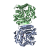
- PDB-5e7f: Complex between lactococcal phage Tuc2009 RBP head domain and a n... -
+ Open data

- Basic information
| Entry | Database: PDB / ID: 5e7f | ||||||
|---|---|---|---|---|---|---|---|
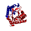
| Title | Complex between lactococcal phage Tuc2009 RBP head domain and a nanobody (L06) | ||||||
 Components Components |
| ||||||
 Keywords Keywords | VIRAL PROTEIN / bacteriophages / Lactococcus lactis / Siphoviridae / receptor binding protein / single-chain nanobody | ||||||
| Function / homology | : / Major structural protein 1, jelly-roll domain / Lower baseplate protein, N-terminal / Lower baseplate protein N-terminal domain / Immunoglobulins / Immunoglobulin-like / Sandwich / Mainly Beta / Major structural protein 1 Function and homology information Function and homology information | ||||||
| Biological species |   Lactococcus phage Tuc2009 (virus) Lactococcus phage Tuc2009 (virus) | ||||||
| Method |  X-RAY DIFFRACTION / SYNCHROTRON / MOLECULAR REPLACEMENT / Resolution: 2.7 Å X-RAY DIFFRACTION / SYNCHROTRON / MOLECULAR REPLACEMENT / Resolution: 2.7 Å | ||||||
 Authors Authors | Legrand, P. / Collins, B. / Blangy, S. / Murphy, J. / Spinelli, S. / Gutierrez, C. / Richet, N. / Kellenberger, C. / Desmyter, A. / Mahony, J. ...Legrand, P. / Collins, B. / Blangy, S. / Murphy, J. / Spinelli, S. / Gutierrez, C. / Richet, N. / Kellenberger, C. / Desmyter, A. / Mahony, J. / van Sinderen, D. / Cambillau, C. | ||||||
 Citation Citation | Journal: Mbio / Year: 2016 Title: The Atomic Structure of the Phage Tuc2009 Baseplate Tripod Suggests that Host Recognition Involves Two Different Carbohydrate Binding Modules. Authors: Legrand, P. / Collins, B. / Blangy, S. / Murphy, J. / Spinelli, S. / Gutierrez, C. / Richet, N. / Kellenberger, C. / Desmyter, A. / Mahony, J. / van Sinderen, D. / Cambillau, C. | ||||||
| History |
|
- Structure visualization
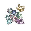
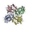
| Structure viewer | Molecule: MolmilJmol/JSmol |
|---|
- Downloads & links
-Download
| PDBx/mmCIF format | 5e7f.cif.gz | 305.4 KB | Display | PDBx/mmCIF format |
|---|---|---|---|---|
| PDB format | pdb5e7f.ent.gz | 251.6 KB | Display | PDB format |
| PDBx/mmJSON format | 5e7f.json.gz | Tree view | PDBx/mmJSON format | |
| Others |  Other downloads Other downloads |
-Validation report
| Summary document | 5e7f_validation.pdf.gz | 466.3 KB | Display | wwPDB validaton report |
|---|---|---|---|---|
| Full document | 5e7f_full_validation.pdf.gz | 474.1 KB | Display | |
| Data in XML | 5e7f_validation.xml.gz | 27.1 KB | Display | |
| Data in CIF | 5e7f_validation.cif.gz | 36.5 KB | Display | |
| Arichive directory | https://data.pdbj.org/pub/pdb/validation_reports/e7/5e7fftp://data.pdbj.org/pub/pdb/validation_reports/e7/5e7f | HTTPS FTP |
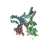
-Related structure data
| Related structure data |  5e7bC  5e7tC  5efbS C: citing same article ( S: Starting model for refinement |
|---|---|
| Similar structure data |


-Links
 PDBj
PDBj

- Assembly
| Deposited unit | 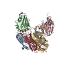
| ||||||||
|---|---|---|---|---|---|---|---|---|---|
| 1 |
| ||||||||
| Unit cell |
|
-Components
| #1: Antibody | Mass: 14164.479 Da / Num. of mol.: 3 Source method: isolated from a genetically manipulated source Details: blood (immunization) / Source: (gene. exp.)  #2: Protein | Mass: 18979.213 Da / Num. of mol.: 3 / Fragment: head domain, UNP residues 1-173 Source method: isolated from a genetically manipulated source Details: Cleaved by trypsin / Source: (gene. exp.) Lactococcus phage Tuc2009 (virus) / Production host: Lactococcus lactis (lactic acid bacteria) / References: UniProt: Q38610#3: Water | ChemComp-HOH / |  Mass: 18.015 Da / Num. of mol.: 40 / Source method: isolated from a natural source / Formula: H2O Mass: 18.015 Da / Num. of mol.: 40 / Source method: isolated from a natural source / Formula: H2OHas protein modification | Y | |
|---|
-Experimental details
-Experiment
| Experiment | Method: X-RAY DIFFRACTION |
|---|
- Sample preparation
| Crystal | Density Matthews: 3.33 Å3/Da / Density % sol: 63.01 % |
|---|---|
| Crystal grow | Temperature: 293 K / Method: vapor diffusion, sitting drop / pH: 7.5 Details: mixing 300 nl of protein (10 mM HEPES, pH 7.5, 150 mM NaCl) with 100 nl precipitant solution (25-30 % PEG4000, 0.2 M Imidazole pH 6.0, or 0.1 M Tris pH 8.0). PH range: 7.5 - 8.0 |
-Data collection
| Diffraction | Mean temperature: 100 K |
|---|---|
| Diffraction source | Source: SYNCHROTRON / Site: SOLEIL  / Beamline: PROXIMA 1 / Wavelength: 0.99 Å / Beamline: PROXIMA 1 / Wavelength: 0.99 Å |
| Detector | Type: PSI PILATUS 6M / Detector: PIXEL / Date: Feb 7, 2015 |
| Radiation | Protocol: SINGLE WAVELENGTH / Monochromatic (M) / Laue (L): M / Scattering type: x-ray |
| Radiation wavelength | Wavelength: 0.99 Å / Relative weight: 1 |
| Reflection | Resolution: 2.7→47.1 Å / Num. obs: 31023 / % possible obs: 99.9 % / Observed criterion σ(F): 0 / Observed criterion σ(I): 0 / Redundancy: 7.2 % / Biso Wilson estimate: 75.17 Å2 / Rmerge(I) obs: 0.146 / Net I/σ(I): 11 |
| Reflection shell | Resolution: 2.7→2.77 Å / Redundancy: 7.1 % / Rmerge(I) obs: 1.6 / Mean I/σ(I) obs: 1.2 / % possible all: 99.9 |
- Processing
| Software |
| |||||||||||||||||||||||||||||||||||||||||||||||||||||||||||||||||||||||||||||||||||||||||||||||||||||||||||||||||||||||||||||||||||||||||||||||||||||||||||||||||||||||||||||||
|---|---|---|---|---|---|---|---|---|---|---|---|---|---|---|---|---|---|---|---|---|---|---|---|---|---|---|---|---|---|---|---|---|---|---|---|---|---|---|---|---|---|---|---|---|---|---|---|---|---|---|---|---|---|---|---|---|---|---|---|---|---|---|---|---|---|---|---|---|---|---|---|---|---|---|---|---|---|---|---|---|---|---|---|---|---|---|---|---|---|---|---|---|---|---|---|---|---|---|---|---|---|---|---|---|---|---|---|---|---|---|---|---|---|---|---|---|---|---|---|---|---|---|---|---|---|---|---|---|---|---|---|---|---|---|---|---|---|---|---|---|---|---|---|---|---|---|---|---|---|---|---|---|---|---|---|---|---|---|---|---|---|---|---|---|---|---|---|---|---|---|---|---|---|---|---|---|
| Refinement | Method to determine structure: MOLECULAR REPLACEMENT Starting model: 5efb Resolution: 2.7→34.8 Å / Cor.coef. Fo:Fc: 0.8883 / Cor.coef. Fo:Fc free: 0.8783 / SU R Cruickshank DPI: 0.464 / Cross valid method: THROUGHOUT / σ(F): 0 / SU R Blow DPI: 0.457 / SU Rfree Blow DPI: 0.24 / SU Rfree Cruickshank DPI: 0.244
| |||||||||||||||||||||||||||||||||||||||||||||||||||||||||||||||||||||||||||||||||||||||||||||||||||||||||||||||||||||||||||||||||||||||||||||||||||||||||||||||||||||||||||||||
| Displacement parameters | Biso mean: 78.75 Å2
| |||||||||||||||||||||||||||||||||||||||||||||||||||||||||||||||||||||||||||||||||||||||||||||||||||||||||||||||||||||||||||||||||||||||||||||||||||||||||||||||||||||||||||||||
| Refine analyze | Luzzati coordinate error obs: 0.395 Å | |||||||||||||||||||||||||||||||||||||||||||||||||||||||||||||||||||||||||||||||||||||||||||||||||||||||||||||||||||||||||||||||||||||||||||||||||||||||||||||||||||||||||||||||
| Refinement step | Cycle: LAST / Resolution: 2.7→34.8 Å
| |||||||||||||||||||||||||||||||||||||||||||||||||||||||||||||||||||||||||||||||||||||||||||||||||||||||||||||||||||||||||||||||||||||||||||||||||||||||||||||||||||||||||||||||
| Refine LS restraints |
| |||||||||||||||||||||||||||||||||||||||||||||||||||||||||||||||||||||||||||||||||||||||||||||||||||||||||||||||||||||||||||||||||||||||||||||||||||||||||||||||||||||||||||||||
| LS refinement shell | Resolution: 2.7→2.79 Å / Total num. of bins used: 16
| |||||||||||||||||||||||||||||||||||||||||||||||||||||||||||||||||||||||||||||||||||||||||||||||||||||||||||||||||||||||||||||||||||||||||||||||||||||||||||||||||||||||||||||||
| Refinement TLS params. | Method: refined / Refine-ID: X-RAY DIFFRACTION
| |||||||||||||||||||||||||||||||||||||||||||||||||||||||||||||||||||||||||||||||||||||||||||||||||||||||||||||||||||||||||||||||||||||||||||||||||||||||||||||||||||||||||||||||
| Refinement TLS group |
|
