 Movie
Movie Controller

[English] 日本語
 Yorodumi
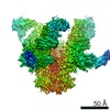
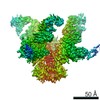
- PDB-5e7t: Structure of the tripod (BppUct-A-L) from the baseplate of bacter... -
+ Open data

- Basic information
| Entry | Database: PDB / ID: 5e7t | ||||||
|---|---|---|---|---|---|---|---|
| Title | Structure of the tripod (BppUct-A-L) from the baseplate of bacteriophage Tuc2009 | ||||||
 Components Components |
| ||||||
 Keywords Keywords | VIRAL PROTEIN / bacteriophages / Lactococcus lactis / Siphoviridae / nanobody / receptor binding protein | ||||||
| Function / homology |  Function and homology information Function and homology information: / Minor structural protein 5-like, C-terminal domain / : / Major structural protein 1, jelly-roll domain / Baseplate upper protein, immunoglobulin like domain / Baseplate upper protein immunoglobulin like domain / BppU, N-terminal / BppU N-terminal domain / Lower baseplate protein, N-terminal / Lower baseplate protein N-terminal domain ...: / Minor structural protein 5-like, C-terminal domain / : / Major structural protein 1, jelly-roll domain / Baseplate upper protein, immunoglobulin like domain / Baseplate upper protein immunoglobulin like domain / BppU, N-terminal / BppU N-terminal domain / Lower baseplate protein, N-terminal / Lower baseplate protein N-terminal domain / : / BppA domain 1 / Fibronectin type III / Fibronectin type III superfamily / Immunoglobulin-like fold Similarity search - Domain/homology | ||||||
| Biological species |  Lactococcus phage Tuc2009 (virus) Lactococcus phage Tuc2009 (virus) | ||||||
| Method |  X-RAY DIFFRACTION / SYNCHROTRON / MOLECULAR REPLACEMENT / Resolution: 2.9 Å X-RAY DIFFRACTION / SYNCHROTRON / MOLECULAR REPLACEMENT / Resolution: 2.9 Å | ||||||
 Authors Authors | Legrand, P. / Collins, B. / Blangy, S. / Murphy, J. / Spinelli, S. / Gutierrez, C. / Richet, N. / Kellenberger, C. / Desmyter, A. / Mahony, J. ...Legrand, P. / Collins, B. / Blangy, S. / Murphy, J. / Spinelli, S. / Gutierrez, C. / Richet, N. / Kellenberger, C. / Desmyter, A. / Mahony, J. / van Sinderen, D. / Cambillau, C. | ||||||
 Citation Citation | Journal: Mbio / Year: 2016 Title: The Atomic Structure of the Phage Tuc2009 Baseplate Tripod Suggests that Host Recognition Involves Two Different Carbohydrate Binding Modules. Authors: Legrand, P. / Collins, B. / Blangy, S. / Murphy, J. / Spinelli, S. / Gutierrez, C. / Richet, N. / Kellenberger, C. / Desmyter, A. / Mahony, J. / van Sinderen, D. / Cambillau, C. | ||||||
| History |
|
- Structure visualization
| Structure viewer | Molecule: MolmilJmol/JSmol |
|---|
- Downloads & links
-Download
| PDBx/mmCIF format | 5e7t.cif.gz | 423.8 KB | Display | PDBx/mmCIF format |
|---|---|---|---|---|
| PDB format | pdb5e7t.ent.gz | 349.3 KB | Display | PDB format |
| PDBx/mmJSON format | 5e7t.json.gz | Tree view | PDBx/mmJSON format | |
| Others |  Other downloads Other downloads |
-Validation report
| Arichive directory | https://data.pdbj.org/pub/pdb/validation_reports/e7/5e7tftp://data.pdbj.org/pub/pdb/validation_reports/e7/5e7t | HTTPS FTP |
|---|
-Related structure data
| Related structure data |  5e7bC  5e7fSC S: Starting model for refinement C: citing same article ( |
|---|---|
| Similar structure data |
-Links
 PDBj
PDBj
- Assembly
| Deposited unit | 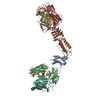
| ||||||||||||
|---|---|---|---|---|---|---|---|---|---|---|---|---|---|
| 1 | 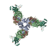
| ||||||||||||
| Unit cell |
| ||||||||||||
| Components on special symmetry positions |
|
-Components
-Minor structural protein ... , 2 types, 2 molecules AB
| #1: Protein | Mass: 14132.773 Da / Num. of mol.: 1 Source method: isolated from a genetically manipulated source Source: (gene. exp.) Lactococcus phage Tuc2009 (virus) / Production host:  Lactococcus lactis (lactic acid bacteria) / References: UniProt: Q9AYV5 Lactococcus lactis (lactic acid bacteria) / References: UniProt: Q9AYV5 |
|---|---|
| #2: Protein | Mass: 31884.508 Da / Num. of mol.: 1 Source method: isolated from a genetically manipulated source Source: (gene. exp.) Lactococcus phage Tuc2009 (virus) / Production host: Lactococcus lactis (lactic acid bacteria) / References: UniProt: Q9AYV4 |
-Major structural protein ... , 2 types, 4 molecules GHIL
| #3: Protein | Mass: 18848.014 Da / Num. of mol.: 1 Source method: isolated from a genetically manipulated source Source: (gene. exp.) Lactococcus phage Tuc2009 (virus) / Production host: Lactococcus lactis (lactic acid bacteria) / References: UniProt: Q38610 |
|---|---|
| #4: Protein | Mass: 18979.213 Da / Num. of mol.: 3 Source method: isolated from a genetically manipulated source Source: (gene. exp.) Lactococcus phage Tuc2009 (virus) / Production host: Lactococcus lactis (lactic acid bacteria) / References: UniProt: Q38610 |
-Non-polymers , 3 types, 67 molecules 


| #5: Chemical | ChemComp-SO4 /  Mass: 96.063 Da / Num. of mol.: 5 / Source method: obtained synthetically / Formula: SO4 Mass: 96.063 Da / Num. of mol.: 5 / Source method: obtained synthetically / Formula: SO4#6: Chemical | ChemComp-CA / |  Mass: 40.078 Da / Num. of mol.: 1 / Source method: obtained synthetically / Formula: Ca Mass: 40.078 Da / Num. of mol.: 1 / Source method: obtained synthetically / Formula: Ca#7: Water | ChemComp-HOH / | Mass: 18.015 Da / Num. of mol.: 61 / Source method: isolated from a natural source / Formula: H2O |
|---|
-Experimental details
-Experiment
| Experiment | Method: X-RAY DIFFRACTION |
|---|
- Sample preparation
| Crystal grow | Temperature: 293 K / Method: vapor diffusion, sitting drop / pH: 7.2 Details: by mixing 300 nl of protein (Na2HPO4, 10 mM; KH2PO4, 1.8 mM [pH7.4]; NaCl, 137 mM; KCl, 2.7 mM) with 100 nl precipitant solution (2 M Ammonium Sulfate, 0.1 M Na Hepes [pH 7]). PH range: 7.0 - 7.4 |
|---|
-Data collection
| Diffraction | Mean temperature: 100 K |
|---|---|
| Diffraction source | Source: SYNCHROTRON / Site: SOLEIL  / Beamline: PROXIMA 1 / Wavelength: 0.97 Å / Beamline: PROXIMA 1 / Wavelength: 0.97 Å |
| Detector | Type: PSI PILATUS 6M / Detector: PIXEL / Date: Jun 6, 2015 |
| Radiation | Protocol: SINGLE WAVELENGTH / Monochromatic (M) / Laue (L): M / Scattering type: x-ray |
| Radiation wavelength | Wavelength: 0.97 Å / Relative weight: 1 |
| Reflection | Resolution: 2.9→34.9 Å / Num. obs: 70164 / % possible obs: 99.8 % / Redundancy: 13.7 % / Biso Wilson estimate: 103.95 Å2 / Rmerge(I) obs: 0.187 / Net I/σ(I): 10.7 |
| Reflection shell | Resolution: 2.9→3 Å / Redundancy: 13.7 % / Rmerge(I) obs: 1.9 / Mean I/σ(I) obs: 1.25 / % possible all: 99.3 |
- Processing
| Software |
| |||||||||||||||||||||||||||||||||||||||||||||||||||||||||||||||||||||||||||||||||||||||||||||||||||||||||||||||||||||||||||||||||||||||||||||||||||||||||||||||||||||||||||||||
|---|---|---|---|---|---|---|---|---|---|---|---|---|---|---|---|---|---|---|---|---|---|---|---|---|---|---|---|---|---|---|---|---|---|---|---|---|---|---|---|---|---|---|---|---|---|---|---|---|---|---|---|---|---|---|---|---|---|---|---|---|---|---|---|---|---|---|---|---|---|---|---|---|---|---|---|---|---|---|---|---|---|---|---|---|---|---|---|---|---|---|---|---|---|---|---|---|---|---|---|---|---|---|---|---|---|---|---|---|---|---|---|---|---|---|---|---|---|---|---|---|---|---|---|---|---|---|---|---|---|---|---|---|---|---|---|---|---|---|---|---|---|---|---|---|---|---|---|---|---|---|---|---|---|---|---|---|---|---|---|---|---|---|---|---|---|---|---|---|---|---|---|---|---|---|---|---|
| Refinement | Method to determine structure: MOLECULAR REPLACEMENT Starting model: 5E7F RBP domain Resolution: 2.9→34.85 Å / Cor.coef. Fo:Fc: 0.9343 / Cor.coef. Fo:Fc free: 0.9233 / SU R Cruickshank DPI: 0.291 / Cross valid method: THROUGHOUT / σ(F): 0 / SU R Blow DPI: 0.278 / SU Rfree Blow DPI: 0.227 / SU Rfree Cruickshank DPI: 0.235
| |||||||||||||||||||||||||||||||||||||||||||||||||||||||||||||||||||||||||||||||||||||||||||||||||||||||||||||||||||||||||||||||||||||||||||||||||||||||||||||||||||||||||||||||
| Displacement parameters | Biso mean: 104.51 Å2
| |||||||||||||||||||||||||||||||||||||||||||||||||||||||||||||||||||||||||||||||||||||||||||||||||||||||||||||||||||||||||||||||||||||||||||||||||||||||||||||||||||||||||||||||
| Refine analyze | Luzzati coordinate error obs: 0.423 Å | |||||||||||||||||||||||||||||||||||||||||||||||||||||||||||||||||||||||||||||||||||||||||||||||||||||||||||||||||||||||||||||||||||||||||||||||||||||||||||||||||||||||||||||||
| Refinement step | Cycle: LAST / Resolution: 2.9→34.85 Å
| |||||||||||||||||||||||||||||||||||||||||||||||||||||||||||||||||||||||||||||||||||||||||||||||||||||||||||||||||||||||||||||||||||||||||||||||||||||||||||||||||||||||||||||||
| Refine LS restraints |
| |||||||||||||||||||||||||||||||||||||||||||||||||||||||||||||||||||||||||||||||||||||||||||||||||||||||||||||||||||||||||||||||||||||||||||||||||||||||||||||||||||||||||||||||
| LS refinement shell | Resolution: 2.9→2.98 Å / Total num. of bins used: 20
| |||||||||||||||||||||||||||||||||||||||||||||||||||||||||||||||||||||||||||||||||||||||||||||||||||||||||||||||||||||||||||||||||||||||||||||||||||||||||||||||||||||||||||||||
| Refinement TLS params. | Method: refined / Refine-ID: X-RAY DIFFRACTION
| |||||||||||||||||||||||||||||||||||||||||||||||||||||||||||||||||||||||||||||||||||||||||||||||||||||||||||||||||||||||||||||||||||||||||||||||||||||||||||||||||||||||||||||||
| Refinement TLS group |
|
