 Movie
Movie Controller


+ Open data

- Basic information
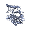
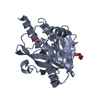
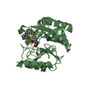
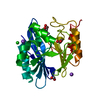
| Entry | Database: PDB / ID: 6lf4 | ||||||
|---|---|---|---|---|---|---|---|
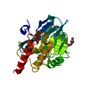
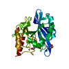
| Title | Crystal structure of VMB-1 bound to hydrolyzed meropenem | ||||||
 Components Components | VMB-1 metallo-beta-lactamase | ||||||
 Keywords Keywords | HYDROLASE / metallo-beta-lactamase / subclasse B1 | ||||||
| Function / homology |  Function and homology information Function and homology informationantibiotic catabolic process / beta-lactamase activity / beta-lactamase / periplasmic space / response to antibiotic / zinc ion binding Similarity search - Function | ||||||
| Biological species |  Vibrio alginolyticus (bacteria) Vibrio alginolyticus (bacteria) | ||||||
| Method |  X-RAY DIFFRACTION / SYNCHROTRON / MOLECULAR REPLACEMENT / Resolution: 2.01 Å X-RAY DIFFRACTION / SYNCHROTRON / MOLECULAR REPLACEMENT / Resolution: 2.01 Å | ||||||
 Authors Authors | Cheng, Q. / Chen, S. | ||||||
 Citation Citation | Journal: To Be Published Title: Crystal structure of VMB-1 bound to hydrolyzed meropenem Authors: Chen, S. / Cheng, Q. | ||||||
| History |
|
- Structure visualization
| Structure viewer | Molecule: MolmilJmol/JSmol |
|---|
- Downloads & links
-Download
| PDBx/mmCIF format | 6lf4.cif.gz | 189.1 KB | Display | PDBx/mmCIF format |
|---|---|---|---|---|
| PDB format | pdb6lf4.ent.gz | 149.1 KB | Display | PDB format |
| PDBx/mmJSON format | 6lf4.json.gz | Tree view | PDBx/mmJSON format | |
| Others |  Other downloads Other downloads |
-Validation report
| Arichive directory | https://data.pdbj.org/pub/pdb/validation_reports/lf/6lf4ftp://data.pdbj.org/pub/pdb/validation_reports/lf/6lf4 | HTTPS FTP |
|---|
-Related structure data
| Related structure data |  6jv4S S: Starting model for refinement |
|---|---|
| Similar structure data |


-Links
 PDBj
PDBj

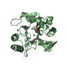
- Assembly
| Deposited unit | 
| ||||||||
|---|---|---|---|---|---|---|---|---|---|
| 1 | 
| ||||||||
| 2 | 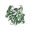
| ||||||||
| 3 | 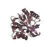
| ||||||||
| 4 | 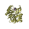
| ||||||||
| Unit cell |
|
-Components
| #1: Protein | Mass: 25705.178 Da / Num. of mol.: 4 Source method: isolated from a genetically manipulated source Source: (gene. exp.) Vibrio alginolyticus (bacteria)Production host: References: UniProt: A0A5Q5ADH9*PLUS #2: Chemical | ChemComp-ZN /   Mass: 65.409 Da / Num. of mol.: 8 / Source method: obtained synthetically / Formula: Zn / Feature type: SUBJECT OF INVESTIGATION Mass: 65.409 Da / Num. of mol.: 8 / Source method: obtained synthetically / Formula: Zn / Feature type: SUBJECT OF INVESTIGATION#3: Chemical | ChemComp-LMP / (   Mass: 401.478 Da / Num. of mol.: 4 / Source method: obtained synthetically / Formula: C17H27N3O6S / Feature type: SUBJECT OF INVESTIGATION Mass: 401.478 Da / Num. of mol.: 4 / Source method: obtained synthetically / Formula: C17H27N3O6S / Feature type: SUBJECT OF INVESTIGATION#4: Water | ChemComp-HOH / |  Mass: 18.015 Da / Num. of mol.: 272 / Source method: isolated from a natural source / Formula: H2O Mass: 18.015 Da / Num. of mol.: 272 / Source method: isolated from a natural source / Formula: H2OHas ligand of interest | Y | |
|---|
-Experimental details
-Experiment
| Experiment | Method: X-RAY DIFFRACTION / Number of used crystals: 1 |
|---|
- Sample preparation
| Crystal | Density Matthews: 2.27 Å3/Da / Density % sol: 45.72 % |
|---|---|
| Crystal grow | Temperature: 289 K / Method: vapor diffusion, hanging drop Details: 0.2 M Ammonium sulfate, 0.1M Bis-Tris pH 5.5, 25% PEG 3350 PH range: 5.4-5.6 |
-Data collection
| Diffraction | Mean temperature: 100 K / Serial crystal experiment: N |
|---|---|
| Diffraction source | Source: SYNCHROTRON / Site: SSRF  / Beamline: BL17U / Wavelength: 0.979191 Å / Beamline: BL17U / Wavelength: 0.979191 Å |
| Detector | Type: DECTRIS EIGER X 16M / Detector: PIXEL / Date: Jun 16, 2019 |
| Radiation | Protocol: SINGLE WAVELENGTH / Monochromatic (M) / Laue (L): M / Scattering type: x-ray |
| Radiation wavelength | Wavelength: 0.979191 Å / Relative weight: 1 |
| Reflection | Resolution: 2.008→77.99 Å / Num. obs: 63155 / % possible obs: 100 % / Redundancy: 13.3 % / Biso Wilson estimate: 32.08 Å2 / CC1/2: 0.92 / Net I/σ(I): 4.8 |
| Reflection shell | Resolution: 2.01→2.12 Å / Redundancy: 13.8 % / Mean I/σ(I) obs: 2.3 / Num. unique obs: 9088 / CC1/2: 0.87 / % possible all: 100 |
- Processing
| Software |
| ||||||||||||||||||||||||||||||||||||||||||||||||||||||||||||
|---|---|---|---|---|---|---|---|---|---|---|---|---|---|---|---|---|---|---|---|---|---|---|---|---|---|---|---|---|---|---|---|---|---|---|---|---|---|---|---|---|---|---|---|---|---|---|---|---|---|---|---|---|---|---|---|---|---|---|---|---|---|
| Refinement | Method to determine structure: MOLECULAR REPLACEMENT Starting model: 6JV4 Resolution: 2.01→77.99 Å / Cor.coef. Fo:Fc: 0.944 / Cor.coef. Fo:Fc free: 0.913 / SU B: 5.369 / SU ML: 0.148 / Cross valid method: THROUGHOUT / σ(F): 0 / ESU R: 0.205 / ESU R Free: 0.181 / Stereochemistry target values: MAXIMUM LIKELIHOOD Details: HYDROGENS HAVE BEEN ADDED IN THE RIDING POSITIONS U VALUES : REFINED INDIVIDUALLY
| ||||||||||||||||||||||||||||||||||||||||||||||||||||||||||||
| Solvent computation | Ion probe radii: 0.8 Å / Shrinkage radii: 0.8 Å / VDW probe radii: 1.2 Å / Solvent model: MASK | ||||||||||||||||||||||||||||||||||||||||||||||||||||||||||||
| Displacement parameters | Biso max: 95.96 Å2 / Biso mean: 35.999 Å2 / Biso min: 19.74 Å2
| ||||||||||||||||||||||||||||||||||||||||||||||||||||||||||||
| Refinement step | Cycle: final / Resolution: 2.01→77.99 Å
| ||||||||||||||||||||||||||||||||||||||||||||||||||||||||||||
| Refine LS restraints |
| ||||||||||||||||||||||||||||||||||||||||||||||||||||||||||||
| LS refinement shell | Resolution: 2.01→2.06 Å / Rfactor Rfree error: 0
|
