- PDB-5e31: 2.3 Angstrom Crystal Structure of the Monomeric Form of Penicilli... -
+
Open data
ID or keywords:
Loading...
-
Basic information
Entry
Database: PDB / ID: 5.0E+31
Title
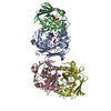
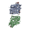
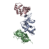
2.3 Angstrom Crystal Structure of the Monomeric Form of Penicillin Binding Protein 2 Prime from Enterococcus faecium.
Components
Penicillin binding protein 2 prime
Keywords
PENICILLIN-BINDING PROTEIN / penicillin binding protein 2 prime / PBP2 / CSGID / Structural Genomics / Center for Structural Genomics of Infectious Diseases
Resolution: 2.3→29.12 Å / Cor.coef. Fo:Fc: 0.946 / Cor.coef. Fo:Fc free: 0.925 / SU B: 13.204 / SU ML: 0.164 / Cross valid method: THROUGHOUT / ESU R: 0.329 / ESU R Free: 0.235 / Stereochemistry target values: MAXIMUM LIKELIHOOD / Details: HYDROGENS HAVE BEEN ADDED IN THE RIDING POSITIONS
Rfactor
Num. reflection
% reflection
Selection details
Rfree
0.24445
1677
5.1 %
RANDOM
Rwork
0.19676
-
-
-
obs
0.19925
30970
98.77 %
-
Solvent computation
Ion probe radii: 0.8 Å / Shrinkage radii: 0.8 Å / VDW probe radii: 1.2 Å / Solvent model: MASK
 Movie
Movie Controller

 Yorodumi
 Open data
 Basic information
 Components
 Keywords
 Function and homology information
 Enterococcus faecium DO (bacteria)
 X-RAY DIFFRACTION /
  Authors
 Citation
 Structure visualization
 Downloads & links
 Other downloads
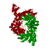


 PDBj
PDBj

 Assembly
 Mass: 18.015 Da / Num. of mol.: 243 / Source method: isolated from a natural source / Formula: H2O
 Sample preparation
 / Beamline: 21-ID-F / Wavelength: 0.97872 Å
 Processing