 Movie
Movie Controller

[English] 日本語
 Yorodumi
- PDB-5dym: Crystal structure of a PadR family transcription regulator from h... -
+ Open data

- Basic information
| Entry | Database: PDB / ID: 5dym | |||||||||
|---|---|---|---|---|---|---|---|---|---|---|
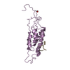
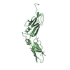
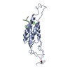
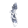
| Title | Crystal structure of a PadR family transcription regulator from hypervirulent Clostridium difficile R20291 - CdPadR_0991 to 1.89 Angstrom resolution | |||||||||
 Components Components | PadR-family transcriptional regulator | |||||||||
 Keywords Keywords | DNA BINDING PROTEIN / PadR / wHTH DNA binding domain / Helix turn helix motifs / transcription regulator / PadR-s2 | |||||||||
| Function / homology | Transcription regulator PadR, N-terminal / Transcriptional regulator PadR-like family / Winged helix-like DNA-binding domain superfamily/Winged helix DNA-binding domain / Arc Repressor Mutant, subunit A / Winged helix DNA-binding domain superfamily / Winged helix-like DNA-binding domain superfamily / Orthogonal Bundle / Mainly Alpha / PadR-family transcriptional regulator Function and homology information Function and homology information | |||||||||
| Biological species |  Peptoclostridium difficile (bacteria) Peptoclostridium difficile (bacteria) | |||||||||
| Method |  X-RAY DIFFRACTION / SYNCHROTRON / MOLECULAR REPLACEMENT / Resolution: 1.894 Å X-RAY DIFFRACTION / SYNCHROTRON / MOLECULAR REPLACEMENT / Resolution: 1.894 Å | |||||||||
 Authors Authors | Isom, C.E. / Karr, E.A. / Menon, S.K. / West, A.H. / Richter-Addo, G.B. | |||||||||
 Citation Citation | Journal: Bmc Microbiol. / Year: 2016 Title: Crystal structure and DNA binding activity of a PadR family transcription regulator from hypervirulent Clostridium difficile R20291. Authors: Isom, C.E. / Menon, S.K. / Thomas, L.M. / West, A.H. / Richter-Addo, G.B. / Karr, E.A. | |||||||||
| History |
|
- Structure visualization
| Structure viewer | Molecule: MolmilJmol/JSmol |
|---|
- Downloads & links
-Download
| PDBx/mmCIF format | 5dym.cif.gz | 34.4 KB | Display | PDBx/mmCIF format |
|---|---|---|---|---|
| PDB format | pdb5dym.ent.gz | 22.1 KB | Display | PDB format |
| PDBx/mmJSON format | 5dym.json.gz | Tree view | PDBx/mmJSON format | |
| Others |  Other downloads Other downloads |
-Validation report
| Arichive directory | https://data.pdbj.org/pub/pdb/validation_reports/dy/5dymftp://data.pdbj.org/pub/pdb/validation_reports/dy/5dym | HTTPS FTP |
|---|
-Related structure data
| Related structure data |  1xmaS S: Starting model for refinement |
|---|---|
| Similar structure data |


-Links
 PDBj
PDBj- Assembly
| Deposited unit | 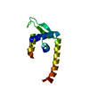
| ||||||||
|---|---|---|---|---|---|---|---|---|---|
| 1 | 
| ||||||||
| Unit cell |
| ||||||||
| Components on special symmetry positions |
|
-Components
| #1: Protein | Mass: 14112.258 Da / Num. of mol.: 1 Source method: isolated from a genetically manipulated source Source: (gene. exp.) Peptoclostridium difficile (strain R20291) (bacteria)Strain: R20291 / Gene: CDR20291_0991 / Plasmid: pQE80 / Production host: |
|---|---|
| #2: Water | ChemComp-HOH /  Mass: 18.015 Da / Num. of mol.: 36 / Source method: isolated from a natural source / Formula: H2O Mass: 18.015 Da / Num. of mol.: 36 / Source method: isolated from a natural source / Formula: H2O |
-Experimental details
-Experiment
| Experiment | Method: X-RAY DIFFRACTION |
|---|
- Sample preparation
| Crystal | Density Matthews: 3.06 Å3/Da / Density % sol: 65.1 % / Description: Square/bipyramidal, clear |
|---|---|
| Crystal grow | Temperature: 298 K / Method: vapor diffusion, hanging drop / pH: 7.5 / Details: 3.1 M NaCl, 100 mM HEPES (pH 7.5) |
-Data collection
| Diffraction | Mean temperature: 100 K |
|---|---|
| Diffraction source | Source: SYNCHROTRON / Site: SSRL  / Beamline: BL14-1 / Wavelength: 1.2 Å / Beamline: BL14-1 / Wavelength: 1.2 Å |
| Detector | Type: MARMOSAIC 325 mm CCD / Detector: CCD / Date: May 14, 2015 |
| Radiation | Protocol: SINGLE WAVELENGTH / Monochromatic (M) / Laue (L): M / Scattering type: x-ray |
| Radiation wavelength | Wavelength: 1.2 Å / Relative weight: 1 |
| Reflection | Resolution: 1.894→34.37 Å / Num. obs: 14302 / % possible obs: 99 % / Redundancy: 2 % / Rmerge(I) obs: 0.018 / Net I/σ(I): 20.27 |
| Reflection shell | Resolution: 1.894→1.962 Å / Redundancy: 2 % / Rmerge(I) obs: 0.3821 / Mean I/σ(I) obs: 2.15 / % possible all: 95.07 |
- Processing
| Software |
| ||||||||||||||||||||||||||||||||||||||||||
|---|---|---|---|---|---|---|---|---|---|---|---|---|---|---|---|---|---|---|---|---|---|---|---|---|---|---|---|---|---|---|---|---|---|---|---|---|---|---|---|---|---|---|---|
| Refinement | Method to determine structure: MOLECULAR REPLACEMENT Starting model: 1XMA Resolution: 1.894→34.369 Å / SU ML: 0.21 / Cross valid method: FREE R-VALUE / σ(F): 1.35 / Phase error: 25.17 / Stereochemistry target values: ML
| ||||||||||||||||||||||||||||||||||||||||||
| Solvent computation | Shrinkage radii: 0.9 Å / VDW probe radii: 1.11 Å / Solvent model: FLAT BULK SOLVENT MODEL | ||||||||||||||||||||||||||||||||||||||||||
| Refinement step | Cycle: LAST / Resolution: 1.894→34.369 Å
| ||||||||||||||||||||||||||||||||||||||||||
| Refine LS restraints |
| ||||||||||||||||||||||||||||||||||||||||||
| LS refinement shell |
|
