 Movie
Movie Controller


+ Open data

- Basic information
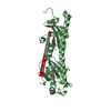
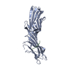
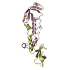
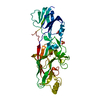
| Entry | Database: PDB / ID: 3ux2 | ||||||
|---|---|---|---|---|---|---|---|
| Title | Crystal Structure of Domain-Swapped Fam96a Major dimer | ||||||
 Components Components | MIP18 family protein FAM96A | ||||||
 Keywords Keywords | IMMUNE SYSTEM / DUF59 / 3D domain swapping / protein-protein interaction / Alpha and beta protein (a+b) / Cytosolic iron-sulfur protein assembly 1 | ||||||
| Function / homology |  Function and homology information Function and homology informationcytosolic [4Fe-4S] assembly targeting complex / iron-sulfur cluster assembly / chromosome segregation / protein maturation / nucleoplasm / metal ion binding / cytoplasm / cytosol Similarity search - Function | ||||||
| Biological species |  Homo sapiens (human) Homo sapiens (human) | ||||||
| Method |  X-RAY DIFFRACTION / SYNCHROTRON / SAD / Resolution: 1.8 Å X-RAY DIFFRACTION / SYNCHROTRON / SAD / Resolution: 1.8 Å | ||||||
 Authors Authors | Chen, K.-E. / Kobe, B. / Martin, J.L. | ||||||
 Citation Citation | Journal: Acta Crystallogr.,Sect.D / Year: 2012 Title: The mammalian DUF59 protein Fam96a forms two distinct types of domain-swapped dimer. Authors: Chen, K.E. / Richards, A.A. / Ariffin, J.K. / Ross, I.L. / Sweet, M.J. / Kellie, S. / Kobe, B. / Martin, J.L. | ||||||
| History |
|
- Structure visualization
| Structure viewer | Molecule: MolmilJmol/JSmol |
|---|
- Downloads & links
-Download
| PDBx/mmCIF format | 3ux2.cif.gz | 62.9 KB | Display | PDBx/mmCIF format |
|---|---|---|---|---|
| PDB format | pdb3ux2.ent.gz | 46.9 KB | Display | PDB format |
| PDBx/mmJSON format | 3ux2.json.gz | Tree view | PDBx/mmJSON format | |
| Others |  Other downloads Other downloads |
-Validation report
| Arichive directory | https://data.pdbj.org/pub/pdb/validation_reports/ux/3ux2ftp://data.pdbj.org/pub/pdb/validation_reports/ux/3ux2 | HTTPS FTP |
|---|
-Related structure data


-Links
 PDBj
PDBj- Assembly
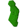
| Deposited unit | 
| ||||||||
|---|---|---|---|---|---|---|---|---|---|
| 1 | 
| ||||||||
| Unit cell |
|
-Components
| #1: Protein | Mass: 15119.002 Da / Num. of mol.: 1 / Fragment: DUF59 Domain, UNP residues 31-157 Source method: isolated from a genetically manipulated source Source: (gene. exp.) Homo sapiens (human) / Gene: FAM96A / Plasmid: pMCSG7 / Production host:  |
|---|---|
| #2: Water | ChemComp-HOH /  Mass: 18.015 Da / Num. of mol.: 128 / Source method: isolated from a natural source / Formula: H2O Mass: 18.015 Da / Num. of mol.: 128 / Source method: isolated from a natural source / Formula: H2O |
| Has protein modification | Y |
-Experimental details
-Experiment
| Experiment | Method: X-RAY DIFFRACTION / Number of used crystals: 1 |
|---|
- Sample preparation
| Crystal | Density Matthews: 1.99 Å3/Da / Density % sol: 38.3 % |
|---|---|
| Crystal grow | Temperature: 278 K / Method: vapor diffusion / pH: 7.5 Details: HEPES, ammonium acetate, PEG 3350, pH 7.5, VAPOR DIFFUSION, temperature 278K |
-Data collection
| Diffraction | Mean temperature: 100 K |
|---|---|
| Diffraction source | Source: SYNCHROTRON / Site: Australian Synchrotron  / Beamline: MX2 / Wavelength: 0.9184 Å / Beamline: MX2 / Wavelength: 0.9184 Å |
| Detector | Type: ADSC QUANTUM 315r / Detector: CCD / Date: Mar 11, 2010 |
| Radiation | Monochromator: Sagitally focused Si / Protocol: SINGLE WAVELENGTH / Scattering type: x-ray |
| Radiation wavelength | Wavelength: 0.9184 Å / Relative weight: 1 |
| Reflection | Resolution: 1.8→58.507 Å / Num. all: 11602 / Num. obs: 11601 / % possible obs: 99.9 % / Observed criterion σ(F): 0 / Observed criterion σ(I): 0 / Redundancy: 13.8 % / Biso Wilson estimate: 14 Å2 / Limit h max: 42 / Limit h min: 0 / Limit k max: 50 / Limit k min: 0 / Limit l max: 19 / Limit l min: 0 / Rmerge(I) obs: 0.06 / Net I/σ(I): 33.9 |
| Reflection scale | Group code: 1 |
| Reflection shell | Resolution: 1.8→1.9 Å / Redundancy: 14.1 % / Rmerge(I) obs: 0.23 / Mean I/σ(I) obs: 15.5 / Num. unique all: 1659 / % possible all: 100 |
-Phasing
| Phasing | Method: SAD |
|---|
- Processing
| Software |
| ||||||||||||||||||||||||||||||||
|---|---|---|---|---|---|---|---|---|---|---|---|---|---|---|---|---|---|---|---|---|---|---|---|---|---|---|---|---|---|---|---|---|---|
| Refinement | Method to determine structure: SAD / Resolution: 1.8→19.541 Å / Occupancy max: 1 / Occupancy min: 0 / FOM work R set: 0.8779 / SU ML: 0.14 / σ(F): 1.97 / Phase error: 18.32 / Stereochemistry target values: ML
| ||||||||||||||||||||||||||||||||
| Solvent computation | Shrinkage radii: 0.65 Å / VDW probe radii: 0.8 Å / Solvent model: FLAT BULK SOLVENT MODEL / Bsol: 42.013 Å2 / ksol: 0.4 e/Å3 | ||||||||||||||||||||||||||||||||
| Displacement parameters | Biso max: 61.22 Å2 / Biso mean: 19.6894 Å2 / Biso min: 4.37 Å2
| ||||||||||||||||||||||||||||||||
| Refine analyze | Luzzati coordinate error obs: 0.14 Å | ||||||||||||||||||||||||||||||||
| Refinement step | Cycle: LAST / Resolution: 1.8→19.541 Å
| ||||||||||||||||||||||||||||||||
| Refine LS restraints |
| ||||||||||||||||||||||||||||||||
| LS refinement shell | Refine-ID: X-RAY DIFFRACTION / Total num. of bins used: 4 / % reflection obs: 100 %
|
