 Movie
Movie Controller

[English] 日本語
 Yorodumi
- PDB-5dxf: Structure of Candida albicans trehalose-6-phosphate phosphatase N... -
+ Open data

- Basic information
| Entry | Database: PDB / ID: 5dxf | ||||||
|---|---|---|---|---|---|---|---|
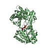
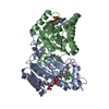
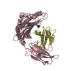
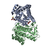
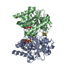
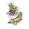
| Title | Structure of Candida albicans trehalose-6-phosphate phosphatase N-terminal domain | ||||||
 Components Components | trehalose-6-phosphate phosphatase | ||||||
 Keywords Keywords | HYDROLASE / trehalose-6-phosphate phosphatase | ||||||
| Function / homology |  Function and homology information Function and homology informationnegative regulation of flocculation / alpha,alpha-trehalose-phosphate synthase complex (UDP-forming) / filamentous growth of a population of unicellular organisms in response to starvation / trehalose-phosphatase activity / trehalose biosynthetic process / filamentous growth / cellular response to osmotic stress / cellular response to starvation / cellular response to heat / cellular response to oxidative stress / metal ion binding Similarity search - Function | ||||||
| Biological species |  Candida albicans (yeast) Candida albicans (yeast) | ||||||
| Method |  X-RAY DIFFRACTION / SYNCHROTRON / MOLECULAR REPLACEMENT / Resolution: 2.563 Å X-RAY DIFFRACTION / SYNCHROTRON / MOLECULAR REPLACEMENT / Resolution: 2.563 Å | ||||||
 Authors Authors | Miao, Y. / Brennan, R.G. | ||||||
| Funding support |  United States, 1items United States, 1items
| ||||||
 Citation Citation | Journal: Proc.Natl.Acad.Sci.USA / Year: 2016 Title: Structures of trehalose-6-phosphate phosphatase from pathogenic fungi reveal the mechanisms of substrate recognition and catalysis. Authors: Miao, Y. / Tenor, J.L. / Toffaletti, D.L. / Washington, E.J. / Liu, J. / Shadrick, W.R. / Schumacher, M.A. / Lee, R.E. / Perfect, J.R. / Brennan, R.G. | ||||||
| History |
|
- Structure visualization
| Structure viewer | Molecule: MolmilJmol/JSmol |
|---|
- Downloads & links
-Download
| PDBx/mmCIF format | 5dxf.cif.gz | 197.4 KB | Display | PDBx/mmCIF format |
|---|---|---|---|---|
| PDB format | pdb5dxf.ent.gz | 156.1 KB | Display | PDB format |
| PDBx/mmJSON format | 5dxf.json.gz | Tree view | PDBx/mmJSON format | |
| Others |  Other downloads Other downloads |
-Validation report
| Summary document | 5dxf_validation.pdf.gz | 444 KB | Display | wwPDB validaton report |
|---|---|---|---|---|
| Full document | 5dxf_full_validation.pdf.gz | 479.3 KB | Display | |
| Data in XML | 5dxf_validation.xml.gz | 36.7 KB | Display | |
| Data in CIF | 5dxf_validation.cif.gz | 50.3 KB | Display | |
| Arichive directory | https://data.pdbj.org/pub/pdb/validation_reports/dx/5dxfftp://data.pdbj.org/pub/pdb/validation_reports/dx/5dxf | HTTPS FTP |
-Related structure data
| Related structure data |  5dx9C  5dxiC  5dxlC 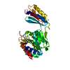 5dxnC 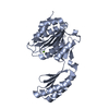 5dxoC 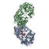 1uquS C: citing same article ( S: Starting model for refinement |
|---|---|
| Similar structure data |


-Links
 PDBj
PDBj- Assembly
| Deposited unit | 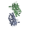
| ||||||||
|---|---|---|---|---|---|---|---|---|---|
| 1 | 
| ||||||||
| 2 | 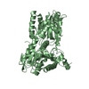
| ||||||||
| Unit cell |
|
-Components
| #1: Protein | Mass: 60756.930 Da / Num. of mol.: 2 / Fragment: N-terminal domain (UNP residues 1-534) Source method: isolated from a genetically manipulated source Source: (gene. exp.) Candida albicans (strain SC5314 / ATCC MYA-2876) (yeast)Strain: SC5314 / ATCC MYA-2876 / Gene: TPS2, CaO19.10556, CaO19.3038, I503_00327, W5Q_00328 / Production host:  References: UniProt: Q5AI14, alpha,alpha-trehalose-phosphate synthase (UDP-forming) #2: Water | ChemComp-HOH / |  Mass: 18.015 Da / Num. of mol.: 98 / Source method: isolated from a natural source / Formula: H2O Mass: 18.015 Da / Num. of mol.: 98 / Source method: isolated from a natural source / Formula: H2O |
|---|
-Experimental details
-Experiment
| Experiment | Method: X-RAY DIFFRACTION / Number of used crystals: 1 |
|---|
- Sample preparation
| Crystal | Density Matthews: 2.6 Å3/Da / Density % sol: 52.68 % |
|---|---|
| Crystal grow | Temperature: 298 K / Method: vapor diffusion, hanging drop / pH: 6.5 / Details: 0.1 M sodium cacodylate, 1.2 M sodium citrate |
-Data collection
| Diffraction | Mean temperature: 200 K | ||||||||||||||||||||||||||||||||||||||||||||||||||||||||||||||||||||||||||||||||||||||||||||||||||||||||||||||||||||||||||||||
|---|---|---|---|---|---|---|---|---|---|---|---|---|---|---|---|---|---|---|---|---|---|---|---|---|---|---|---|---|---|---|---|---|---|---|---|---|---|---|---|---|---|---|---|---|---|---|---|---|---|---|---|---|---|---|---|---|---|---|---|---|---|---|---|---|---|---|---|---|---|---|---|---|---|---|---|---|---|---|---|---|---|---|---|---|---|---|---|---|---|---|---|---|---|---|---|---|---|---|---|---|---|---|---|---|---|---|---|---|---|---|---|---|---|---|---|---|---|---|---|---|---|---|---|---|---|---|---|
| Diffraction source | Source: SYNCHROTRON / Site: APS / Beamline: 22-ID / Wavelength: 1 Å | ||||||||||||||||||||||||||||||||||||||||||||||||||||||||||||||||||||||||||||||||||||||||||||||||||||||||||||||||||||||||||||||
| Detector | Type: MARMOSAIC 300 mm CCD / Detector: CCD / Date: Oct 7, 2013 | ||||||||||||||||||||||||||||||||||||||||||||||||||||||||||||||||||||||||||||||||||||||||||||||||||||||||||||||||||||||||||||||
| Radiation | Monochromator: double crystal Si(111) / Protocol: SINGLE WAVELENGTH / Monochromatic (M) / Laue (L): M / Scattering type: x-ray | ||||||||||||||||||||||||||||||||||||||||||||||||||||||||||||||||||||||||||||||||||||||||||||||||||||||||||||||||||||||||||||||
| Radiation wavelength | Wavelength: 1 Å / Relative weight: 1 | ||||||||||||||||||||||||||||||||||||||||||||||||||||||||||||||||||||||||||||||||||||||||||||||||||||||||||||||||||||||||||||||
| Reflection | Resolution: 2.56→50 Å / Num. obs: 42110 / % possible obs: 97.6 % / Redundancy: 6.1 % / Biso Wilson estimate: 50.86 Å2 / Rmerge(I) obs: 0.098 / Χ2: 1.3 / Net I/av σ(I): 21.437 / Net I/σ(I): 9.2 / Num. measured all: 258931 | ||||||||||||||||||||||||||||||||||||||||||||||||||||||||||||||||||||||||||||||||||||||||||||||||||||||||||||||||||||||||||||||
| Reflection shell | Diffraction-ID: 1 / Rejects: _
|
- Processing
| Software |
| |||||||||||||||||||||||||||||||||||||||||||||||||||||||||||||||||||||||||||||||||||||||||||||||||||||||||
|---|---|---|---|---|---|---|---|---|---|---|---|---|---|---|---|---|---|---|---|---|---|---|---|---|---|---|---|---|---|---|---|---|---|---|---|---|---|---|---|---|---|---|---|---|---|---|---|---|---|---|---|---|---|---|---|---|---|---|---|---|---|---|---|---|---|---|---|---|---|---|---|---|---|---|---|---|---|---|---|---|---|---|---|---|---|---|---|---|---|---|---|---|---|---|---|---|---|---|---|---|---|---|---|---|---|---|
| Refinement | Method to determine structure: MOLECULAR REPLACEMENT Starting model: PDB entry 1UQU Resolution: 2.563→48.868 Å / FOM work R set: 0.7251 / SU ML: 0.38 / Cross valid method: FREE R-VALUE / σ(F): 1.34 / Phase error: 30.04 / Stereochemistry target values: ML
| |||||||||||||||||||||||||||||||||||||||||||||||||||||||||||||||||||||||||||||||||||||||||||||||||||||||||
| Solvent computation | Shrinkage radii: 0.9 Å / VDW probe radii: 1.11 Å / Solvent model: FLAT BULK SOLVENT MODEL | |||||||||||||||||||||||||||||||||||||||||||||||||||||||||||||||||||||||||||||||||||||||||||||||||||||||||
| Displacement parameters | Biso max: 169.39 Å2 / Biso mean: 61.69 Å2 / Biso min: 16.66 Å2 | |||||||||||||||||||||||||||||||||||||||||||||||||||||||||||||||||||||||||||||||||||||||||||||||||||||||||
| Refinement step | Cycle: final / Resolution: 2.563→48.868 Å
| |||||||||||||||||||||||||||||||||||||||||||||||||||||||||||||||||||||||||||||||||||||||||||||||||||||||||
| Refine LS restraints |
| |||||||||||||||||||||||||||||||||||||||||||||||||||||||||||||||||||||||||||||||||||||||||||||||||||||||||
| LS refinement shell | Refine-ID: X-RAY DIFFRACTION / Total num. of bins used: 14
|
