 Movie
Movie Controller


+ Open data

- Basic information
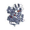
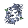
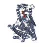
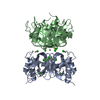
| Entry | Database: PDB / ID: 5dvm | ||||||
|---|---|---|---|---|---|---|---|
| Title | Fc Design 20.8.37 B chain homodimer E357D/S364R/Y407A | ||||||
 Components Components |
| ||||||
 Keywords Keywords | IMMUNE SYSTEM / head-to-tail homodimer / Immunoglobulin / Fc | ||||||
| Function / homology |  Function and homology information Function and homology informationcomplement-dependent cytotoxicity / antibody-dependent cellular cytotoxicity / Fc-gamma receptor I complex binding / immunoglobulin complex, circulating / Classical antibody-mediated complement activation / immunoglobulin receptor binding / IgG immunoglobulin complex / Initial triggering of complement / FCGR activation / complement activation, classical pathway ...complement-dependent cytotoxicity / antibody-dependent cellular cytotoxicity / Fc-gamma receptor I complex binding / immunoglobulin complex, circulating / Classical antibody-mediated complement activation / immunoglobulin receptor binding / IgG immunoglobulin complex / Initial triggering of complement / FCGR activation / complement activation, classical pathway / Role of phospholipids in phagocytosis / antigen binding / FCGR3A-mediated IL10 synthesis / Regulation of Complement cascade / B cell receptor signaling pathway / FCGR3A-mediated phagocytosis / Regulation of actin dynamics for phagocytic cup formation / antibacterial humoral response / Interleukin-4 and Interleukin-13 signaling / blood microparticle / adaptive immune response / : / extracellular exosome / extracellular region / plasma membrane Similarity search - Function | ||||||
| Biological species |  Homo sapiens (human) Homo sapiens (human)synthetic construct (others) | ||||||
| Method |  X-RAY DIFFRACTION / SYNCHROTRON / Resolution: 2.95 Å X-RAY DIFFRACTION / SYNCHROTRON / Resolution: 2.95 Å | ||||||
| Model details | Design XXX | ||||||
 Authors Authors | Atwell, S. / Leaver-Fay, A. / Froning, K.J. / Aldaz, H. / Pustilnik, A. / Lu, F. / Huang, F. / Yuan, R. / Dhanani, S.H. / Chamberlain, A.K. ...Atwell, S. / Leaver-Fay, A. / Froning, K.J. / Aldaz, H. / Pustilnik, A. / Lu, F. / Huang, F. / Yuan, R. / Dhanani, S.H. / Chamberlain, A.K. / Fitchett, J.R. / Gutierrez, B. / Hendle, J. / Demarest, S.J. / Kuhlman, B. | ||||||
 Citation Citation | Journal: Structure / Year: 2016 Title: Computationally Designed Bispecific Antibodies using Negative State Repertoires. Authors: Leaver-Fay, A. / Froning, K.J. / Atwell, S. / Aldaz, H. / Pustilnik, A. / Lu, F. / Huang, F. / Yuan, R. / Hassanali, S. / Chamberlain, A.K. / Fitchett, J.R. / Demarest, S.J. / Kuhlman, B. | ||||||
| History |
|
- Structure visualization
| Structure viewer | Molecule: MolmilJmol/JSmol |
|---|
- Downloads & links
-Download
| PDBx/mmCIF format | 5dvm.cif.gz | 58.6 KB | Display | PDBx/mmCIF format |
|---|---|---|---|---|
| PDB format | pdb5dvm.ent.gz | 40.7 KB | Display | PDB format |
| PDBx/mmJSON format | 5dvm.json.gz | Tree view | PDBx/mmJSON format | |
| Others |  Other downloads Other downloads |
-Validation report
| Arichive directory | https://data.pdbj.org/pub/pdb/validation_reports/dv/5dvmftp://data.pdbj.org/pub/pdb/validation_reports/dv/5dvm | HTTPS FTP |
|---|
-Related structure data
| Related structure data | 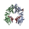 5di8C 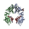 5dj0C 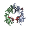 5dj2C 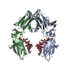 5dj6C  5dj8C  5djaC  5djcC  5djdC  5djxC  5djyC  5djzC  5dk0C  5dk2C  5dvkC  5dvlC  5dvnC  5dvoC C: citing same article ( |
|---|---|
| Similar structure data |


-Links
 PDBj
PDBj


- Assembly
| Deposited unit | 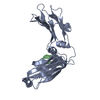
| ||||||||
|---|---|---|---|---|---|---|---|---|---|
| 1 | 
| ||||||||
| Unit cell |
|
-Components
| #1: Protein | Mass: 25577.012 Da / Num. of mol.: 1 / Fragment: UNP residues 104-330 / Mutation: E357D, S364R, Y407A Source method: isolated from a genetically manipulated source Details: transient expression / Source: (gene. exp.) Homo sapiens (human) / Gene: IGHG1 / Cell line (production host): HEK293 / Production host: Homo sapiens (human) / References: UniProt: P01857 |
|---|---|
| #2: Protein/peptide | Mass: 1533.749 Da / Num. of mol.: 1 / Source method: obtained synthetically / Source: (synth.) synthetic construct (others) |
| #3: Water | ChemComp-HOH /  Mass: 18.015 Da / Num. of mol.: 7 / Source method: isolated from a natural source / Formula: H2O Mass: 18.015 Da / Num. of mol.: 7 / Source method: isolated from a natural source / Formula: H2O |
| Has protein modification | Y |
-Experimental details
-Experiment
| Experiment | Method: X-RAY DIFFRACTION / Number of used crystals: 1 |
|---|
- Sample preparation
| Crystal | Density Matthews: 3.76 Å3/Da / Density % sol: 67.27 % |
|---|---|
| Crystal grow | Temperature: 294 K / Method: vapor diffusion Details: 100mM Sodium Cacodylate pH 6.5 + 20% PEG 8K + 200mM Magnesium Acetate tetrahydrate |
-Data collection
| Diffraction | Mean temperature: 193 K | ||||||||||||||||||||||||||||||||||||||||||||||||||||||||||||||||||||||||||||||||||||||||||||||||||||||||||||||
|---|---|---|---|---|---|---|---|---|---|---|---|---|---|---|---|---|---|---|---|---|---|---|---|---|---|---|---|---|---|---|---|---|---|---|---|---|---|---|---|---|---|---|---|---|---|---|---|---|---|---|---|---|---|---|---|---|---|---|---|---|---|---|---|---|---|---|---|---|---|---|---|---|---|---|---|---|---|---|---|---|---|---|---|---|---|---|---|---|---|---|---|---|---|---|---|---|---|---|---|---|---|---|---|---|---|---|---|---|---|---|---|
| Diffraction source | Source: SYNCHROTRON / Site: APS  / Beamline: 31-ID / Wavelength: 0.97931 Å / Beamline: 31-ID / Wavelength: 0.97931 Å | ||||||||||||||||||||||||||||||||||||||||||||||||||||||||||||||||||||||||||||||||||||||||||||||||||||||||||||||
| Detector | Type: RAYONIX MX-225 / Detector: CCD / Date: Jun 2, 2014 / Details: Diamond (111) | ||||||||||||||||||||||||||||||||||||||||||||||||||||||||||||||||||||||||||||||||||||||||||||||||||||||||||||||
| Radiation | Protocol: SINGLE WAVELENGTH / Monochromatic (M) / Laue (L): M / Scattering type: x-ray | ||||||||||||||||||||||||||||||||||||||||||||||||||||||||||||||||||||||||||||||||||||||||||||||||||||||||||||||
| Radiation wavelength | Wavelength: 0.97931 Å / Relative weight: 1 | ||||||||||||||||||||||||||||||||||||||||||||||||||||||||||||||||||||||||||||||||||||||||||||||||||||||||||||||
| Reflection | Resolution: 2.95→30 Å / Num. obs: 8056 / % possible obs: 98.7 % / Redundancy: 5.7 % / Rpim(I) all: 0.062 / Rrim(I) all: 0.162 / Rsym value: 0.136 / Net I/av σ(I): 4.279 / Net I/σ(I): 7.9 / Num. measured all: 45883 | ||||||||||||||||||||||||||||||||||||||||||||||||||||||||||||||||||||||||||||||||||||||||||||||||||||||||||||||
| Reflection shell | Diffraction-ID: 1 / Rejects: _
|
- Processing
| Software |
| |||||||||||||||||||||||||||||||||||||||||||||
|---|---|---|---|---|---|---|---|---|---|---|---|---|---|---|---|---|---|---|---|---|---|---|---|---|---|---|---|---|---|---|---|---|---|---|---|---|---|---|---|---|---|---|---|---|---|---|
| Refinement | Resolution: 2.95→30 Å / Cor.coef. Fo:Fc: 0.93 / Cor.coef. Fo:Fc free: 0.886 / SU B: 20.583 / SU ML: 0.36 / Cross valid method: THROUGHOUT / σ(F): 0 / ESU R: 0.948 / ESU R Free: 0.409 / Stereochemistry target values: MAXIMUM LIKELIHOOD / Details: U VALUES : REFINED INDIVIDUALLY
| |||||||||||||||||||||||||||||||||||||||||||||
| Solvent computation | Ion probe radii: 0.8 Å / Shrinkage radii: 0.8 Å / VDW probe radii: 1.2 Å / Solvent model: MASK | |||||||||||||||||||||||||||||||||||||||||||||
| Displacement parameters | Biso max: 146.5 Å2 / Biso mean: 69.424 Å2 / Biso min: 32.96 Å2
| |||||||||||||||||||||||||||||||||||||||||||||
| Refinement step | Cycle: final / Resolution: 2.95→30 Å
| |||||||||||||||||||||||||||||||||||||||||||||
| Refine LS restraints |
| |||||||||||||||||||||||||||||||||||||||||||||
| LS refinement shell | Resolution: 2.95→3.026 Å / Total num. of bins used: 20
|
