 Movie
Movie Controller


+ Open data

- Basic information
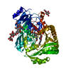
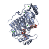
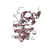
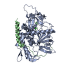
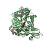
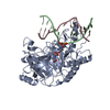
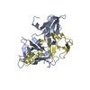
| Entry | Database: PDB / ID: 4quv | ||||||
|---|---|---|---|---|---|---|---|
| Title | Structure of an integral membrane delta(14)-sterol reductase | ||||||
 Components Components | Delta(14)-sterol reductase | ||||||
 Keywords Keywords | Oxidoreductase / Membrane protein / Cholesterol Biosynthesis | ||||||
| Function / homology |  Function and homology information Function and homology informationDelta14-sterol reductase / Delta14-sterol reductase activity / sterol biosynthetic process / ergosterol biosynthetic process / NADP binding / plasma membrane Similarity search - Function | ||||||
| Biological species |  Methylomicrobium alcaliphilum (bacteria) Methylomicrobium alcaliphilum (bacteria) | ||||||
| Method |  X-RAY DIFFRACTION / SYNCHROTRON / SAD / Resolution: 2.743 Å X-RAY DIFFRACTION / SYNCHROTRON / SAD / Resolution: 2.743 Å | ||||||
 Authors Authors | Li, X. / Blobel, G. | ||||||
 Citation Citation | Journal: Nature / Year: 2015 Title: Structure of an integral membrane sterol reductase from Methylomicrobium alcaliphilum. Authors: Li, X. / Roberti, R. / Blobel, G. | ||||||
| History |
|
- Structure visualization
| Structure viewer | Molecule: MolmilJmol/JSmol |
|---|
- Downloads & links
-Download
| PDBx/mmCIF format | 4quv.cif.gz | 337.7 KB | Display | PDBx/mmCIF format |
|---|---|---|---|---|
| PDB format | pdb4quv.ent.gz | 277.7 KB | Display | PDB format |
| PDBx/mmJSON format | 4quv.json.gz | Tree view | PDBx/mmJSON format | |
| Others |  Other downloads Other downloads |
-Validation report
| Arichive directory | https://data.pdbj.org/pub/pdb/validation_reports/qu/4quvftp://data.pdbj.org/pub/pdb/validation_reports/qu/4quv | HTTPS FTP |
|---|
-Related structure data
| Similar structure data |
|---|


-Links
 PDBj
PDBj

- Assembly
| Deposited unit | 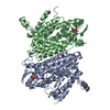
| ||||||||
|---|---|---|---|---|---|---|---|---|---|
| 1 | 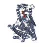
| ||||||||
| 2 | 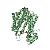
| ||||||||
| 3 |
| ||||||||
| Unit cell |
|
-Components
| #1: Protein | Mass: 49466.613 Da / Num. of mol.: 2 Source method: isolated from a genetically manipulated source Source: (gene. exp.) Methylomicrobium alcaliphilum (bacteria)Strain: DSM 19304 / NCIMB 14124 / VKM B-2133 / 20Z / Gene: erg, MEALZ_1312 / Production host: #2: Chemical |   Mass: 745.421 Da / Num. of mol.: 2 / Source method: obtained synthetically / Formula: C21H30N7O17P3 Mass: 745.421 Da / Num. of mol.: 2 / Source method: obtained synthetically / Formula: C21H30N7O17P3 |
|---|
-Experimental details
-Experiment
| Experiment | Method: X-RAY DIFFRACTION / Number of used crystals: 1 |
|---|
- Sample preparation
| Crystal | Density Matthews: 4.08 Å3/Da / Density % sol: 69.88 % |
|---|---|
| Crystal grow | Temperature: 293 K / Method: vapor diffusion, hanging drop / pH: 7 Details: 0.1 M Tris, 0.2 M NH4Ac, 30% (v/v) Pentaerythritol ethoxylate (15/4 EO/OH), pH 7, VAPOR DIFFUSION, HANGING DROP, temperature 293K |
-Data collection
| Diffraction | Mean temperature: 100 K |
|---|---|
| Diffraction source | Source: SYNCHROTRON / Site: NSLS  / Beamline: X29A / Wavelength: 1.075 Å / Beamline: X29A / Wavelength: 1.075 Å |
| Detector | Type: ADSC QUANTUM 315r / Detector: CCD / Date: Oct 13, 2013 |
| Radiation | Protocol: SINGLE WAVELENGTH / Monochromatic (M) / Laue (L): M / Scattering type: x-ray |
| Radiation wavelength | Wavelength: 1.075 Å / Relative weight: 1 |
| Reflection | Resolution: 2.74→37.25 Å / Num. all: 63092 / Num. obs: 30541 / % possible obs: 74.8 % / Redundancy: 2.1 % / Rmerge(I) obs: 0.041 / Rsym value: 0.058 / Net I/σ(I): 21.1 |
| Reflection shell | Resolution: 2.74→2.84 Å / Redundancy: 1.9 % / Rmerge(I) obs: 0.415 / Mean I/σ(I) obs: 1.4 / Rsym value: 0.514 / % possible all: 28.5 |
- Processing
| Software |
| ||||||||||||||||||||||||||||||||||||||||||||||||||||||||||||||||||||||||||||||||||||
|---|---|---|---|---|---|---|---|---|---|---|---|---|---|---|---|---|---|---|---|---|---|---|---|---|---|---|---|---|---|---|---|---|---|---|---|---|---|---|---|---|---|---|---|---|---|---|---|---|---|---|---|---|---|---|---|---|---|---|---|---|---|---|---|---|---|---|---|---|---|---|---|---|---|---|---|---|---|---|---|---|---|---|---|---|---|
| Refinement | Method to determine structure: SAD / Resolution: 2.743→37.255 Å / SU ML: 0.43 / σ(F): 1.96 / Phase error: 37.46 / Stereochemistry target values: ML
| ||||||||||||||||||||||||||||||||||||||||||||||||||||||||||||||||||||||||||||||||||||
| Solvent computation | Shrinkage radii: 0.9 Å / VDW probe radii: 1.11 Å / Solvent model: FLAT BULK SOLVENT MODEL | ||||||||||||||||||||||||||||||||||||||||||||||||||||||||||||||||||||||||||||||||||||
| Refinement step | Cycle: LAST / Resolution: 2.743→37.255 Å
| ||||||||||||||||||||||||||||||||||||||||||||||||||||||||||||||||||||||||||||||||||||
| Refine LS restraints |
| ||||||||||||||||||||||||||||||||||||||||||||||||||||||||||||||||||||||||||||||||||||
| LS refinement shell |
| ||||||||||||||||||||||||||||||||||||||||||||||||||||||||||||||||||||||||||||||||||||
| Refinement TLS params. | Method: refined / Refine-ID: X-RAY DIFFRACTION
| ||||||||||||||||||||||||||||||||||||||||||||||||||||||||||||||||||||||||||||||||||||
| Refinement TLS group |
|
