 Movie
Movie Controller

[English] 日本語
 Yorodumi
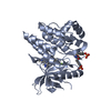
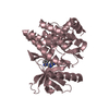
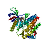
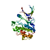
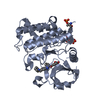
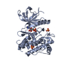
- PDB-5dt6: Crystal structure of the Drosophila GluR1A ligand binding domain ... -
+ Open data

- Basic information
| Entry | Database: PDB / ID: 5dt6 | ||||||
|---|---|---|---|---|---|---|---|
| Title | Crystal structure of the Drosophila GluR1A ligand binding domain complex with glutamate | ||||||
 Components Components | Glutamate receptor 1 | ||||||
 Keywords Keywords | MEMBRANE PROTEIN | ||||||
| Function / homology |  Function and homology information Function and homology informationActivation of AMPA receptors / Trafficking of GluR2-containing AMPA receptors / Synaptic adhesion-like molecules / Unblocking of NMDA receptors, glutamate binding and activation / glutamate receptor activity / kainate selective glutamate receptor activity / ionotropic glutamate receptor complex / monoatomic cation transport / synaptic transmission, glutamatergic / transmitter-gated monoatomic ion channel activity involved in regulation of postsynaptic membrane potential ...Activation of AMPA receptors / Trafficking of GluR2-containing AMPA receptors / Synaptic adhesion-like molecules / Unblocking of NMDA receptors, glutamate binding and activation / glutamate receptor activity / kainate selective glutamate receptor activity / ionotropic glutamate receptor complex / monoatomic cation transport / synaptic transmission, glutamatergic / transmitter-gated monoatomic ion channel activity involved in regulation of postsynaptic membrane potential / postsynaptic density membrane / modulation of chemical synaptic transmission / plasma membrane Similarity search - Function | ||||||
| Biological species |  | ||||||
| Method |  X-RAY DIFFRACTION / SYNCHROTRON / MOLECULAR REPLACEMENT / Resolution: 1.598 Å X-RAY DIFFRACTION / SYNCHROTRON / MOLECULAR REPLACEMENT / Resolution: 1.598 Å | ||||||
 Authors Authors | Dharkar, P. / Mayer, M.L. | ||||||
 Citation Citation | Journal: Neuron / Year: 2016 Title: Novel Functional Properties of Drosophila CNS Glutamate Receptors. Authors: Li, Y. / Dharkar, P. / Han, T.H. / Serpe, M. / Lee, C.H. / Mayer, M.L. | ||||||
| History |
|
- Structure visualization
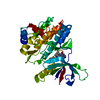
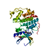
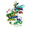
| Structure viewer | Molecule: MolmilJmol/JSmol |
|---|
- Downloads & links
-Download
| PDBx/mmCIF format | 5dt6.cif.gz | 173.1 KB | Display | PDBx/mmCIF format |
|---|---|---|---|---|
| PDB format | pdb5dt6.ent.gz | 139.5 KB | Display | PDB format |
| PDBx/mmJSON format | 5dt6.json.gz | Tree view | PDBx/mmJSON format | |
| Others |  Other downloads Other downloads |
-Validation report
| Arichive directory | https://data.pdbj.org/pub/pdb/validation_reports/dt/5dt6ftp://data.pdbj.org/pub/pdb/validation_reports/dt/5dt6 | HTTPS FTP |
|---|
-Related structure data
| Related structure data | 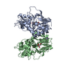 5dtbC  5ehmC  5ehsC  5ictC  1s50S C: citing same article ( S: Starting model for refinement |
|---|---|
| Similar structure data |

-Links
 PDBj
PDBj


- Assembly
| Deposited unit | 
| ||||||||
|---|---|---|---|---|---|---|---|---|---|
| 1 |
| ||||||||
| Unit cell |
|
-Components
| #1: Protein | Mass: 30566.738 Da / Num. of mol.: 1 / Fragment: unp residues 474-594; unp residues 739-880 Source method: isolated from a genetically manipulated source Source: (gene. exp.)  | ||||
|---|---|---|---|---|---|
| #2: Chemical | ChemComp-GLU /   Type: L-peptide linking / Mass: 147.129 Da / Num. of mol.: 1 / Source method: obtained synthetically / Formula: C5H9NO4 Type: L-peptide linking / Mass: 147.129 Da / Num. of mol.: 1 / Source method: obtained synthetically / Formula: C5H9NO4 | ||||
| #3: Chemical |   Mass: 92.094 Da / Num. of mol.: 2 / Source method: obtained synthetically / Formula: C3H8O3 Mass: 92.094 Da / Num. of mol.: 2 / Source method: obtained synthetically / Formula: C3H8O3#4: Water | ChemComp-HOH / |  Mass: 18.015 Da / Num. of mol.: 245 / Source method: isolated from a natural source / Formula: H2O Mass: 18.015 Da / Num. of mol.: 245 / Source method: isolated from a natural source / Formula: H2OHas protein modification | Y | |
-Experimental details
-Experiment
| Experiment | Method: X-RAY DIFFRACTION / Number of used crystals: 1 |
|---|
- Sample preparation
| Crystal | Density Matthews: 2.55 Å3/Da / Density % sol: 51.82 % |
|---|---|
| Crystal grow | Temperature: 293 K / Method: vapor diffusion, hanging drop / pH: 8.5 Details: Sample buffer 150 NaCl, 10 Tris pH 8.5, 2 glutamate, 1 EDTA, 10% glycerol Reservoir 13% PEG 8K, 100 Tris pH 8.5 |
-Data collection
| Diffraction | Mean temperature: 100 K |
|---|---|
| Diffraction source | Source: SYNCHROTRON / Site: APS  / Beamline: 22-ID / Wavelength: 1 Å / Beamline: 22-ID / Wavelength: 1 Å |
| Detector | Type: MARMOSAIC 300 mm CCD / Detector: CCD / Date: Oct 23, 2014 |
| Radiation | Protocol: SINGLE WAVELENGTH / Monochromatic (M) / Laue (L): M / Scattering type: x-ray |
| Radiation wavelength | Wavelength: 1 Å / Relative weight: 1 |
| Reflection | Resolution: 1.54→50 Å / Num. obs: 46081 / % possible obs: 98.4 % / Redundancy: 5.5 % / Rmerge(I) obs: 0.038 / Net I/σ(I): 26.4 |
| Reflection shell | Resolution: 1.54→1.57 Å / Redundancy: 3.9 % / Rmerge(I) obs: 0.63 / Mean I/σ(I) obs: 1.42 / % possible all: 95.2 |
- Processing
| Software |
| ||||||||||||||||||||||||||||||||||||||||||||||||||||||||||||||||||||||||||||||||||||||||||||||||||||||||||||||||||||||||||||||||||||||||||||||||||||||||||||||||||||||||||||||||||||||||||||||||||||||||
|---|---|---|---|---|---|---|---|---|---|---|---|---|---|---|---|---|---|---|---|---|---|---|---|---|---|---|---|---|---|---|---|---|---|---|---|---|---|---|---|---|---|---|---|---|---|---|---|---|---|---|---|---|---|---|---|---|---|---|---|---|---|---|---|---|---|---|---|---|---|---|---|---|---|---|---|---|---|---|---|---|---|---|---|---|---|---|---|---|---|---|---|---|---|---|---|---|---|---|---|---|---|---|---|---|---|---|---|---|---|---|---|---|---|---|---|---|---|---|---|---|---|---|---|---|---|---|---|---|---|---|---|---|---|---|---|---|---|---|---|---|---|---|---|---|---|---|---|---|---|---|---|---|---|---|---|---|---|---|---|---|---|---|---|---|---|---|---|---|---|---|---|---|---|---|---|---|---|---|---|---|---|---|---|---|---|---|---|---|---|---|---|---|---|---|---|---|---|---|---|---|---|
| Refinement | Method to determine structure: MOLECULAR REPLACEMENT Starting model: 1S50 Resolution: 1.598→29.176 Å / SU ML: 0.13 / Cross valid method: THROUGHOUT / σ(F): 1.34 / Phase error: 15.95 / Stereochemistry target values: ML
| ||||||||||||||||||||||||||||||||||||||||||||||||||||||||||||||||||||||||||||||||||||||||||||||||||||||||||||||||||||||||||||||||||||||||||||||||||||||||||||||||||||||||||||||||||||||||||||||||||||||||
| Solvent computation | Shrinkage radii: 0.9 Å / VDW probe radii: 1.11 Å / Solvent model: FLAT BULK SOLVENT MODEL | ||||||||||||||||||||||||||||||||||||||||||||||||||||||||||||||||||||||||||||||||||||||||||||||||||||||||||||||||||||||||||||||||||||||||||||||||||||||||||||||||||||||||||||||||||||||||||||||||||||||||
| Refinement step | Cycle: LAST / Resolution: 1.598→29.176 Å
| ||||||||||||||||||||||||||||||||||||||||||||||||||||||||||||||||||||||||||||||||||||||||||||||||||||||||||||||||||||||||||||||||||||||||||||||||||||||||||||||||||||||||||||||||||||||||||||||||||||||||
| Refine LS restraints |
| ||||||||||||||||||||||||||||||||||||||||||||||||||||||||||||||||||||||||||||||||||||||||||||||||||||||||||||||||||||||||||||||||||||||||||||||||||||||||||||||||||||||||||||||||||||||||||||||||||||||||
| LS refinement shell |
| ||||||||||||||||||||||||||||||||||||||||||||||||||||||||||||||||||||||||||||||||||||||||||||||||||||||||||||||||||||||||||||||||||||||||||||||||||||||||||||||||||||||||||||||||||||||||||||||||||||||||
| Refinement TLS params. | Method: refined / Refine-ID: X-RAY DIFFRACTION
| ||||||||||||||||||||||||||||||||||||||||||||||||||||||||||||||||||||||||||||||||||||||||||||||||||||||||||||||||||||||||||||||||||||||||||||||||||||||||||||||||||||||||||||||||||||||||||||||||||||||||
| Refinement TLS group |
|
