 Movie
Movie Controller

[English] 日本語
 Yorodumi
- PDB-5dmm: Crystal Structure of the Homocysteine Methyltransferase MmuM from... -
+ Open data

- Basic information
| Entry | Database: PDB / ID: 5dmm | ||||||
|---|---|---|---|---|---|---|---|
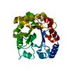
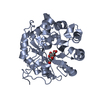
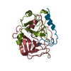
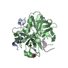
| Title | Crystal Structure of the Homocysteine Methyltransferase MmuM from Escherichia coli, Metallated form | ||||||
 Components Components | Homocysteine S-methyltransferase | ||||||
 Keywords Keywords | TRANSFERASE / Homocysteine Methyltransferase | ||||||
| Function / homology |  Function and homology information Function and homology informationhomocysteine S-methyltransferase / S-adenosylmethionine-homocysteine S-methyltransferase activity / S-methylmethionine metabolic process / S-methylmethionine cycle / S-methylmethionine-homocysteine S-methyltransferase activity / methionine biosynthetic process / methylation / DNA damage response / zinc ion binding Similarity search - Function | ||||||
| Biological species |  | ||||||
| Method |  X-RAY DIFFRACTION / SYNCHROTRON / MOLECULAR REPLACEMENT / Resolution: 1.779 Å X-RAY DIFFRACTION / SYNCHROTRON / MOLECULAR REPLACEMENT / Resolution: 1.779 Å | ||||||
 Authors Authors | Li, K. / Li, G. / Bradbury, L.M.T. / Andrew, H.D. / Bruner, S.D. | ||||||
 Citation Citation | Journal: Biochem.J. / Year: 2016 Title: Crystal structure of the homocysteine methyltransferase MmuM from Escherichia coli. Authors: Li, K. / Li, G. / Bradbury, L.M. / Hanson, A.D. / Bruner, S.D. | ||||||
| History |
|
- Structure visualization
| Structure viewer | Molecule: MolmilJmol/JSmol |
|---|
- Downloads & links
-Download
| PDBx/mmCIF format | 5dmm.cif.gz | 73.2 KB | Display | PDBx/mmCIF format |
|---|---|---|---|---|
| PDB format | pdb5dmm.ent.gz | 52.6 KB | Display | PDB format |
| PDBx/mmJSON format | 5dmm.json.gz | Tree view | PDBx/mmJSON format | |
| Others |  Other downloads Other downloads |
-Validation report
| Summary document | 5dmm_validation.pdf.gz | 452.9 KB | Display | wwPDB validaton report |
|---|---|---|---|---|
| Full document | 5dmm_full_validation.pdf.gz | 453.1 KB | Display | |
| Data in XML | 5dmm_validation.xml.gz | 14 KB | Display | |
| Data in CIF | 5dmm_validation.cif.gz | 20 KB | Display | |
| Arichive directory | https://data.pdbj.org/pub/pdb/validation_reports/dm/5dmmftp://data.pdbj.org/pub/pdb/validation_reports/dm/5dmm | HTTPS FTP |
-Related structure data
| Related structure data | 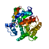 5dmlC  5dmnC  1q7mS C: citing same article ( S: Starting model for refinement |
|---|---|
| Similar structure data |


-Links
 PDBj
PDBj


- Assembly
| Deposited unit | 
| |||||||||
|---|---|---|---|---|---|---|---|---|---|---|
| 1 |
| |||||||||
| Unit cell |
| |||||||||
| Components on special symmetry positions |
|
-Components
| #1: Protein | Mass: 33457.754 Da / Num. of mol.: 1 Source method: isolated from a genetically manipulated source Source: (gene. exp.) Strain: K12 / Gene: mmuM, yagD, b0261, JW0253 / Production host: References: UniProt: Q47690, homocysteine S-methyltransferase | ||||||
|---|---|---|---|---|---|---|---|
| #2: Chemical |   Mass: 65.409 Da / Num. of mol.: 3 / Source method: obtained synthetically / Formula: Zn Mass: 65.409 Da / Num. of mol.: 3 / Source method: obtained synthetically / Formula: Zn#3: Chemical | ChemComp-BME / |   Mass: 78.133 Da / Num. of mol.: 1 / Source method: obtained synthetically / Formula: C2H6OS Mass: 78.133 Da / Num. of mol.: 1 / Source method: obtained synthetically / Formula: C2H6OS#4: Chemical | ChemComp-HCS / |   Type: L-peptide linking / Mass: 135.185 Da / Num. of mol.: 1 / Source method: obtained synthetically / Formula: C4H9NO2S Type: L-peptide linking / Mass: 135.185 Da / Num. of mol.: 1 / Source method: obtained synthetically / Formula: C4H9NO2S#5: Water | ChemComp-HOH / |  Mass: 18.015 Da / Num. of mol.: 180 / Source method: isolated from a natural source / Formula: H2O Mass: 18.015 Da / Num. of mol.: 180 / Source method: isolated from a natural source / Formula: H2O |
-Experimental details
-Experiment
| Experiment | Method: X-RAY DIFFRACTION |
|---|
- Sample preparation
| Crystal | Density Matthews: 2.41 Å3/Da / Density % sol: 48.93 % / Description: rod-shaped |
|---|---|
| Crystal grow | Temperature: 298 K / Method: vapor diffusion, hanging drop / pH: 4.6 Details: 1.6 M ammonium sulphate, 10% v/v glycerol and 0.1 M sodium acetate pH 4.8 PH range: 4.8 |
-Data collection
| Diffraction | Mean temperature: 100 K |
|---|---|
| Diffraction source | Source: SYNCHROTRON / Site: APS  / Beamline: 21-ID-G / Wavelength: 0.9787 Å / Beamline: 21-ID-G / Wavelength: 0.9787 Å |
| Detector | Type: MARMOSAIC 300 mm CCD / Detector: CCD / Date: Jul 22, 2015 |
| Radiation | Monochromator: Diamond [111] / Protocol: SINGLE WAVELENGTH / Monochromatic (M) / Laue (L): M / Scattering type: x-ray |
| Radiation wavelength | Wavelength: 0.9787 Å / Relative weight: 1 |
| Reflection | Resolution: 1.779→24.23 Å / Num. obs: 13523 / % possible obs: 93.4 % / Redundancy: 5.2 % / Rmerge(I) obs: 0.093 / Rsym value: 0.104 / Net I/σ(I): 14.86 |
| Reflection shell | Resolution: 1.779→1.84 Å / Redundancy: 5 % / Rmerge(I) obs: 0.66 / Mean I/σ(I) obs: 2.49 / % possible all: 95.36 |
- Processing
| Software |
| |||||||||||||||||||||||||||||||||||||||||||||||||||||||||||||||||||||||||||||
|---|---|---|---|---|---|---|---|---|---|---|---|---|---|---|---|---|---|---|---|---|---|---|---|---|---|---|---|---|---|---|---|---|---|---|---|---|---|---|---|---|---|---|---|---|---|---|---|---|---|---|---|---|---|---|---|---|---|---|---|---|---|---|---|---|---|---|---|---|---|---|---|---|---|---|---|---|---|---|
| Refinement | Method to determine structure: MOLECULAR REPLACEMENT Starting model: 1q7m Resolution: 1.779→24.225 Å / SU ML: 0.16 / Cross valid method: FREE R-VALUE / σ(F): 1.34 / Phase error: 20.37 / Stereochemistry target values: ML
| |||||||||||||||||||||||||||||||||||||||||||||||||||||||||||||||||||||||||||||
| Solvent computation | Shrinkage radii: 0.9 Å / VDW probe radii: 1.11 Å / Solvent model: FLAT BULK SOLVENT MODEL | |||||||||||||||||||||||||||||||||||||||||||||||||||||||||||||||||||||||||||||
| Refinement step | Cycle: LAST / Resolution: 1.779→24.225 Å
| |||||||||||||||||||||||||||||||||||||||||||||||||||||||||||||||||||||||||||||
| Refine LS restraints |
| |||||||||||||||||||||||||||||||||||||||||||||||||||||||||||||||||||||||||||||
| LS refinement shell |
|
