 Movie
Movie Controller


+ Open data

- Basic information
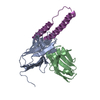
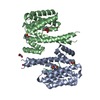
| Entry | Database: PDB / ID: 5dhz | ||||||||||||
|---|---|---|---|---|---|---|---|---|---|---|---|---|---|
| Title | HIV-1 Rev NTD dimers with variable crossing angles | ||||||||||||
 Components Components |
| ||||||||||||
 Keywords Keywords | IMMUNE SYSTEM / HIV / helical hairpin / nuclear export / RNA-binding | ||||||||||||
| Function / homology |  Function and homology information Function and homology informationhost cell nucleolus / viral process / mRNA transport / protein export from nucleus / host cell cytoplasm / DNA-binding transcription factor activity / RNA binding Similarity search - Function | ||||||||||||
| Biological species |    Human immunodeficiency virus 1 Human immunodeficiency virus 1 | ||||||||||||
| Method |  X-RAY DIFFRACTION / SYNCHROTRON / MOLECULAR REPLACEMENT / Resolution: 4.3 Å X-RAY DIFFRACTION / SYNCHROTRON / MOLECULAR REPLACEMENT / Resolution: 4.3 Å | ||||||||||||
 Authors Authors | DiMattia, M.A. / Watts, N.R. / Wingfield, P.T. / Grimes, J.M. / Stuart, D.I. / Steven, A.C. | ||||||||||||
| Funding support |  United States, United States,  United Kingdom, 3items United Kingdom, 3items
| ||||||||||||
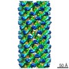
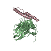
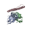
 Citation Citation | Journal: Structure / Year: 2016 Title: The Structure of HIV-1 Rev Filaments Suggests a Bilateral Model for Rev-RRE Assembly. Authors: Michael A DiMattia / Norman R Watts / Naiqian Cheng / Rick Huang / J Bernard Heymann / Jonathan M Grimes / Paul T Wingfield / David I Stuart / Alasdair C Steven / Abstract: HIV-1 Rev protein mediates the nuclear export of viral RNA genomes. To do so, Rev oligomerizes cooperatively onto an RNA motif, the Rev response element (RRE), forming a complex that engages with the ...HIV-1 Rev protein mediates the nuclear export of viral RNA genomes. To do so, Rev oligomerizes cooperatively onto an RNA motif, the Rev response element (RRE), forming a complex that engages with the host nuclear export machinery. To better understand Rev oligomerization, we determined four crystal structures of Rev N-terminal domain dimers, which show that they can pivot about their dyad axis, giving crossing angles of 90° to 140°. In parallel, we performed cryoelectron microscopy of helical Rev filaments. Filaments vary from 11 to 15 nm in width, reflecting variations in dimer crossing angle. These structures contain additional density, indicating that C-terminal domains become partially ordered in the context of filaments. This conformational variability may be exploited in the assembly of RRE/Rev complexes. Our data also revealed a third interface between Revs, which offers an explanation for how the arrangement of Rev subunits adapts to the "A"-shaped architecture of the RRE in export-active complexes. | ||||||||||||
| History |
|
- Structure visualization
| Structure viewer | Molecule: MolmilJmol/JSmol |
|---|
- Downloads & links
-Download
| PDBx/mmCIF format | 5dhz.cif.gz | 66.9 KB | Display | PDBx/mmCIF format |
|---|---|---|---|---|
| PDB format | pdb5dhz.ent.gz | 49 KB | Display | PDB format |
| PDBx/mmJSON format | 5dhz.json.gz | Tree view | PDBx/mmJSON format | |
| Others |  Other downloads Other downloads |
-Validation report
| Arichive directory | https://data.pdbj.org/pub/pdb/validation_reports/dh/5dhzftp://data.pdbj.org/pub/pdb/validation_reports/dh/5dhz | HTTPS FTP |
|---|
-Related structure data


-Links
 PDBj
PDBj


- Assembly
| Deposited unit | 
| ||||||||
|---|---|---|---|---|---|---|---|---|---|
| 1 |
| ||||||||
| Unit cell |
|
-Components
| #1: Antibody | Mass: 12579.941 Da / Num. of mol.: 1 Source method: isolated from a genetically manipulated source Source: (gene. exp.)  |
|---|---|
| #2: Antibody | Mass: 11656.958 Da / Num. of mol.: 1 Source method: isolated from a genetically manipulated source Source: (gene. exp.) |
| #3: Protein | Mass: 7750.793 Da / Num. of mol.: 1 Source method: isolated from a genetically manipulated source Source: (gene. exp.) Human immunodeficiency virus 1 / Gene: rev / Production host: |
| Has protein modification | Y |
-Experimental details
-Experiment
| Experiment | Method: X-RAY DIFFRACTION |
|---|
- Sample preparation
| Crystal | Density Matthews: 2.81 Å3/Da / Density % sol: 56.26 % |
|---|---|
| Crystal grow | Temperature: 293 K / Method: vapor diffusion, sitting drop Details: Crystals of scFv-Rev were initially grown in 20% PEG 3350, a variety of salts (200 mM sodium sulfate, sodium bromide, or ammonium phosphate dibasic), and pH ranging from 6.5-8.5 PH range: 6.5 - 8.5 |
-Data collection
| Diffraction | Mean temperature: 80 K |
|---|---|
| Diffraction source | Source: SYNCHROTRON / Site: ESRF  / Beamline: ID23-2 / Wavelength: 0.8726 Å / Beamline: ID23-2 / Wavelength: 0.8726 Å |
| Detector | Type: MARMOSAIC 225 mm CCD / Detector: CCD / Date: Jul 1, 2010 |
| Radiation | Protocol: SINGLE WAVELENGTH / Monochromatic (M) / Laue (L): M / Scattering type: x-ray |
| Radiation wavelength | Wavelength: 0.8726 Å / Relative weight: 1 |
| Reflection | Resolution: 4.3→50 Å / Num. obs: 2588 / % possible obs: 98.3 % / Redundancy: 3.1 % / Net I/σ(I): 20.4 |
| Reflection shell | Resolution: 4.3→4.4 Å / Redundancy: 2.8 % / Mean I/σ(I) obs: 1.1 / % possible all: 98.6 |
- Processing
| Software |
| ||||||||||||||||
|---|---|---|---|---|---|---|---|---|---|---|---|---|---|---|---|---|---|
| Refinement | Method to determine structure: MOLECULAR REPLACEMENT / Resolution: 4.3→50 Å / Cross valid method: FREE R-VALUE
| ||||||||||||||||
| Refinement step | Cycle: LAST / Resolution: 4.3→50 Å
|
