 Movie
Movie Controller

[English] 日本語
 Yorodumi
- PDB-5d92: Structure of a phosphatidylinositolphosphate (PIP) synthase from ... -
+ Open data

- Basic information
| Entry | Database: PDB / ID: 5d92 | ||||||
|---|---|---|---|---|---|---|---|
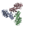
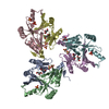
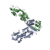
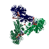
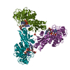
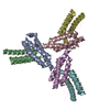
| Title | Structure of a phosphatidylinositolphosphate (PIP) synthase from Renibacterium Salmoninarum | ||||||
 Components Components | AF2299 protein,Phosphatidylinositol synthase | ||||||
 Keywords Keywords | MEMBRANE PROTEIN / Enzyme / lipid biosynthesis / phosphatidylinositol | ||||||
| Function / homology |  Function and homology information Function and homology informationTransferases; Transferring phosphorus-containing groups; Transferases for other substituted phosphate groups / phosphotransferase activity, for other substituted phosphate groups / phospholipid biosynthetic process / nucleotide binding / magnesium ion binding / membrane / metal ion binding / plasma membrane Similarity search - Function | ||||||
| Biological species |   Archaeoglobus fulgidus (archaea) Archaeoglobus fulgidus (archaea) Renibacterium salmoninarum (bacteria) Renibacterium salmoninarum (bacteria) | ||||||
| Method |  X-RAY DIFFRACTION / SYNCHROTRON / MOLECULAR REPLACEMENT / Resolution: 3.62 Å X-RAY DIFFRACTION / SYNCHROTRON / MOLECULAR REPLACEMENT / Resolution: 3.62 Å | ||||||
 Authors Authors | Clarke, O.B. / Tomasek, D.T. / Jorge, C.D. / Belcher Dufrisne, M. / Kim, M. / Banerjee, S. / Rajashankar, K.R. / Hendrickson, W.A. / Santos, H. / Mancia, F. | ||||||
 Citation Citation | Journal: Nat Commun / Year: 2015 Title: Structural basis for phosphatidylinositol-phosphate biosynthesis. Authors: Clarke, O.B. / Tomasek, D. / Jorge, C.D. / Dufrisne, M.B. / Kim, M. / Banerjee, S. / Rajashankar, K.R. / Shapiro, L. / Hendrickson, W.A. / Santos, H. / Mancia, F. | ||||||
| History |
|
- Structure visualization
| Structure viewer | Molecule: MolmilJmol/JSmol |
|---|
- Downloads & links
-Download
| PDBx/mmCIF format | 5d92.cif.gz | 287.8 KB | Display | PDBx/mmCIF format |
|---|---|---|---|---|
| PDB format | pdb5d92.ent.gz | 226.8 KB | Display | PDB format |
| PDBx/mmJSON format | 5d92.json.gz | Tree view | PDBx/mmJSON format | |
| Others |  Other downloads Other downloads |
-Validation report
| Arichive directory | https://data.pdbj.org/pub/pdb/validation_reports/d9/5d92ftp://data.pdbj.org/pub/pdb/validation_reports/d9/5d92 | HTTPS FTP |
|---|
-Related structure data
| Related structure data |  5d91SC S: Starting model for refinement C: citing same article ( |
|---|---|
| Similar structure data |


-Links
 PDBj
PDBj- Assembly
| Deposited unit | 
| |||||||||||||||||||||||||||||||||||
|---|---|---|---|---|---|---|---|---|---|---|---|---|---|---|---|---|---|---|---|---|---|---|---|---|---|---|---|---|---|---|---|---|---|---|---|---|
| 1 | 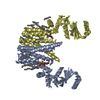
| |||||||||||||||||||||||||||||||||||
| 2 | 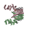
| |||||||||||||||||||||||||||||||||||
| Unit cell |
| |||||||||||||||||||||||||||||||||||
| Noncrystallographic symmetry (NCS) | NCS domain:
NCS domain segments: Component-ID: 1 / Ens-ID: 1 / Beg auth comp-ID: MET / Beg label comp-ID: MET / End auth comp-ID: ALA / End label comp-ID: ALA / Auth seq-ID: -137 - 204 / Label seq-ID: 1 - 342
|
-Components
| #1: Protein | Mass: 37597.832 Da / Num. of mol.: 4 Source method: isolated from a genetically manipulated source Source: (gene. exp.) Archaeoglobus fulgidus (archaea), (gene. exp.) Renibacterium salmoninarum (bacteria)Strain: ATCC 49558 / VC-16 / DSM 4304 / JCM 9628 / NBRC 100126, ATCC 33209 / DSM 20767 / JCM 11484 / NBRC 15589 / NCIMB 2235 Gene: AF_2299, RSal33209_2010 / Production host: #2: Chemical | ChemComp-8K6 /   Mass: 254.494 Da / Num. of mol.: 33 / Source method: obtained synthetically / Formula: C18H38 Mass: 254.494 Da / Num. of mol.: 33 / Source method: obtained synthetically / Formula: C18H38#3: Chemical | ChemComp-MG /   Mass: 24.305 Da / Num. of mol.: 8 / Source method: obtained synthetically / Formula: Mg Mass: 24.305 Da / Num. of mol.: 8 / Source method: obtained synthetically / Formula: Mg#4: Chemical | ChemComp-58A /   Mass: 1006.147 Da / Num. of mol.: 4 / Source method: obtained synthetically / Formula: C48H85N3O15P2 Mass: 1006.147 Da / Num. of mol.: 4 / Source method: obtained synthetically / Formula: C48H85N3O15P2 |
|---|
-Experimental details
-Experiment
| Experiment | Method: X-RAY DIFFRACTION / Number of used crystals: 1 |
|---|
- Sample preparation
| Crystal | Density Matthews: 3.09 Å3/Da / Density % sol: 60.24 % |
|---|---|
| Crystal grow | Temperature: 293 K / Method: lipidic cubic phase Details: 30% (v/v) PEG 300, 0.1 M MES pH 6.0, 0.1 M sodium chloride, 0.1 M magnesium chloride (precipitant); Concentrated protein was mixed with molten monoolein in a 1:1.5 (w/w) ratio of protein: ...Details: 30% (v/v) PEG 300, 0.1 M MES pH 6.0, 0.1 M sodium chloride, 0.1 M magnesium chloride (precipitant); Concentrated protein was mixed with molten monoolein in a 1:1.5 (w/w) ratio of protein:lipid using coupled syringes. A Mosquito LCP (TTP Labtech) robot was used to dispense a typical volume of 50-75 nL of protein/lipid mixture onto a 96-well glass sandwich plate, which was covered with 750 nL precipitant solution. Monoolein was doped with 2% CDP-DAG. |
-Data collection
| Diffraction | Mean temperature: 100 K | |||||||||||||||||||||||||||
|---|---|---|---|---|---|---|---|---|---|---|---|---|---|---|---|---|---|---|---|---|---|---|---|---|---|---|---|---|
| Diffraction source | Source: SYNCHROTRON / Site: APS  / Beamline: 24-ID-C / Wavelength: 0.9791 Å / Beamline: 24-ID-C / Wavelength: 0.9791 Å | |||||||||||||||||||||||||||
| Detector | Type: PSI PILATUS 6M / Detector: PIXEL / Date: Jun 9, 2014 | |||||||||||||||||||||||||||
| Radiation | Protocol: SINGLE WAVELENGTH / Monochromatic (M) / Laue (L): M / Scattering type: x-ray | |||||||||||||||||||||||||||
| Radiation wavelength | Wavelength: 0.9791 Å / Relative weight: 1 | |||||||||||||||||||||||||||
| Reflection | Resolution: 3.62→166.97 Å / % possible obs: 98.9 % / Redundancy: 3.7 % / Biso Wilson estimate: 79.39 Å2 / CC1/2: 0.991 / Rmerge(I) obs: 0.252 / Rpim(I) all: 0.15 / Net I/σ(I): 5 / Num. measured all: 78622 | |||||||||||||||||||||||||||
| Reflection shell | Diffraction-ID: 1 / Rejects: _
|
- Processing
| Software |
| ||||||||||||||||||||||||||||||||||||||||||||||||||||||||
|---|---|---|---|---|---|---|---|---|---|---|---|---|---|---|---|---|---|---|---|---|---|---|---|---|---|---|---|---|---|---|---|---|---|---|---|---|---|---|---|---|---|---|---|---|---|---|---|---|---|---|---|---|---|---|---|---|---|
| Refinement | Method to determine structure: MOLECULAR REPLACEMENT Starting model: 5D91 Resolution: 3.62→166.97 Å / SU ML: 0.57 / Cross valid method: FREE R-VALUE / σ(F): 1.34 / Phase error: 32.89 / Stereochemistry target values: ML
| ||||||||||||||||||||||||||||||||||||||||||||||||||||||||
| Solvent computation | Shrinkage radii: 0.8 Å / VDW probe radii: 1.1 Å / Solvent model: FLAT BULK SOLVENT MODEL | ||||||||||||||||||||||||||||||||||||||||||||||||||||||||
| Displacement parameters | Biso max: 185.71 Å2 / Biso mean: 76.5321 Å2 / Biso min: 26.35 Å2 | ||||||||||||||||||||||||||||||||||||||||||||||||||||||||
| Refinement step | Cycle: final / Resolution: 3.62→166.97 Å
| ||||||||||||||||||||||||||||||||||||||||||||||||||||||||
| Refine LS restraints |
| ||||||||||||||||||||||||||||||||||||||||||||||||||||||||
| Refine LS restraints NCS |
| ||||||||||||||||||||||||||||||||||||||||||||||||||||||||
| LS refinement shell | Refine-ID: X-RAY DIFFRACTION / Total num. of bins used: 7
|
