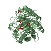
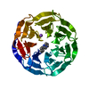
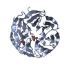
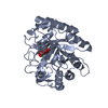
[English] 日本語
Yorodumi
- PDB-5d8z: Structrue of a lucidum protein -

+
Open data


ID or keywords:

Loading...

-
Basic information

Entry
Database: PDB / ID: 5d8z
TitleStructrue of a lucidum protein
ComponentsendoglucanaseCellulase
KeywordsHYDROLASE / GH5 family / Ganoderma lucidum / endoglucanase
Function / homology
Function and homology information


hydrolase activity, hydrolyzing O-glycosyl compounds / carbohydrate metabolic process
Similarity search - Function
Glycoside hydrolase, family 5, conserved site / Glycosyl hydrolases family 5 signature. / Glycoside hydrolase, family 5 / Cellulase (glycosyl hydrolase family 5) / Glycosidases / Glycoside hydrolase superfamily / TIM Barrel / Alpha-Beta Barrel / Alpha Beta
Similarity search - Domain/homology
beta-cellobiose / Endoglucanase
Similarity search - Component
Biological speciesGanoderma lucidum (fungus)
MethodX-RAY DIFFRACTION / SYNCHROTRON / MOLECULAR REPLACEMENT / Resolution: 2.7 Å
AuthorsGuo, R. / Li, Q. / Shang, N. / Liu, G. / Ko, T.P. / Chen, C.C. / Liu, W.
CitationJournal: Enzyme.Microb.Technol. / Year: 2016
Title: Functional and structural analyses of a 1,4-beta-endoglucanase from Ganoderma lucidum.
Authors: Liu, G. / Li, Q. / Shang, N. / Huang, J.W. / Ko, T.P. / Liu, W. / Zheng, Y. / Han, X. / Chen, Y. / Chen, C.C. / Jin, J. / Guo, R.T.
History
DepositionAug 18, 2015Deposition site: RCSB / Processing site: PDBJ
Revision 1.0Jun 29, 2016Provider: repository / Type: Initial release
Revision 1.1May 23, 2018Group: Data collection / Derived calculations / Category: diffrn_source / pdbx_struct_oper_list
Item: _diffrn_source.pdbx_synchrotron_beamline / _diffrn_source.pdbx_synchrotron_site ..._diffrn_source.pdbx_synchrotron_beamline / _diffrn_source.pdbx_synchrotron_site / _diffrn_source.source / _pdbx_struct_oper_list.symmetry_operation
Revision 2.0Jul 29, 2020Group: Atomic model / Data collection ...Atomic model / Data collection / Derived calculations / Non-polymer description / Structure summary
Category: atom_site / chem_comp ...atom_site / chem_comp / entity / entity_name_com / pdbx_branch_scheme / pdbx_chem_comp_identifier / pdbx_entity_branch / pdbx_entity_branch_descriptor / pdbx_entity_branch_link / pdbx_entity_branch_list / pdbx_entity_nonpoly / pdbx_molecule_features / pdbx_nonpoly_scheme / struct_conn / struct_conn_type / struct_site / struct_site_gen
Item: _atom_site.B_iso_or_equiv / _atom_site.Cartn_x ..._atom_site.B_iso_or_equiv / _atom_site.Cartn_x / _atom_site.Cartn_y / _atom_site.Cartn_z / _atom_site.auth_asym_id / _atom_site.auth_atom_id / _atom_site.auth_comp_id / _atom_site.auth_seq_id / _atom_site.label_atom_id / _atom_site.label_comp_id / _atom_site.type_symbol / _chem_comp.formula / _chem_comp.formula_weight / _chem_comp.id / _chem_comp.name / _chem_comp.type / _entity.formula_weight / _entity.pdbx_description / _entity.type
Description: Carbohydrate remediation / Provider: repository / Type: Remediation
Revision 2.1Nov 8, 2023Group: Data collection / Database references ...Data collection / Database references / Refinement description / Structure summary
Category: chem_comp / chem_comp_atom ...chem_comp / chem_comp_atom / chem_comp_bond / database_2 / pdbx_initial_refinement_model
Item: _chem_comp.pdbx_synonyms / _database_2.pdbx_DOI / _database_2.pdbx_database_accession

-
Structure visualization

Structure viewerMolecule:
MolmilJmol/JSmol

Downloads & links

-
Assembly

Deposited unit
A: endoglucanase
B: endoglucanase
hetero molecules


Theoretical massNumber of molelcules
Total (without water)74,1614
Polymers73,4762
Non-polymers6852
Water1,31573
1
A: endoglucanase
hetero molecules


Theoretical massNumber of molelcules
Total (without water)37,0802
Polymers36,7381
Non-polymers3421
Water181
TypeNameSymmetry operationNumber
identity operation1_555x,y,z1
2
B: endoglucanase
hetero molecules


Theoretical massNumber of molelcules
Total (without water)37,0802
Polymers36,7381
Non-polymers3421
Water181
TypeNameSymmetry operationNumber
identity operation1_555x,y,z1
Unit cell
Length a, b, c (Å)84.426, 236.399, 83.585
Angle α, β, γ (deg.)90.00, 90.00, 90.00
Int Tables number20
Space group name H-MC2221

-
Components

#1: Protein endoglucanase / Cellulase


Mass: 36738.000 Da / Num. of mol.: 2
Source method: isolated from a genetically manipulated source
Source: (gene. exp.) Ganoderma lucidum (fungus) / Production host: Escherichia coli (E. coli) / References: UniProt: A0A1A9TAJ1*PLUS
#2: Polysaccharide beta-D-glucopyranose-(1-4)-beta-D-glucopyranose / beta-cellobiose


Type: oligosaccharide, Oligosaccharide / Class: Metabolism / Mass: 342.297 Da / Num. of mol.: 2
Source method: isolated from a genetically manipulated source
Details: oligosaccharide / References: beta-cellobiose
DescriptorTypeProgram
DGlcpb1-4DGlcpb1-ROHGlycam Condensed SequenceGMML 1.0
WURCS=2.0/1,2,1/[a2122h-1b_1-5]/1-1/a4-b1WURCSPDB2Glycan 1.1.0
[][b-D-Glcp]{[(4+1)][b-D-Glcp]{}}LINUCSPDB-CARE
#3: Water ChemComp-HOH / water / Water


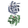
Mass: 18.015 Da / Num. of mol.: 73 / Source method: isolated from a natural source / Formula: H2O

-
Experimental details

-
Experiment

ExperimentMethod: X-RAY DIFFRACTION

-
Sample preparation

CrystalDensity Matthews: 2.84 Å3/Da / Density % sol: 56.66 %
Crystal growTemperature: 298 K / Method: vapor diffusion, sitting drop / pH: 5.5
Details: 0.2M Magnesium chloride hexahydrate, 0.1M BIS-TRIS pH 5.5, 25% w/v Polyethylene glycol 3350

-
Data collection

DiffractionMean temperature: 100 K
Diffraction sourceSource: SYNCHROTRON / Site: NSRRC / Beamline: BL13B1 / Wavelength: 0.97622 Å
DetectorType: ADSC QUANTUM 315 / Detector: CCD / Date: May 10, 2015
RadiationProtocol: SINGLE WAVELENGTH / Monochromatic (M) / Laue (L): M / Scattering type: x-ray
Radiation wavelengthWavelength: 0.97622 Å / Relative weight: 1
ReflectionResolution: 2.7→25 Å / Num. obs: 21889 / % possible obs: 93.4 % / Redundancy: 7.8 % / Net I/σ(I): 20.41

-
Processing

Software
NameVersionClassification
REFMAC5.8.0049refinement
HKL-2000data reduction
HKL-2000data scaling
PHASERphasing
RefinementMethod to determine structure: MOLECULAR REPLACEMENT
Starting model: 3QR3
Resolution: 2.7→25 Å / Cor.coef. Fo:Fc: 0.95 / Cor.coef. Fo:Fc free: 0.92 / SU B: 11.887 / SU ML: 0.24 / Cross valid method: THROUGHOUT / ESU R: 1.261 / ESU R Free: 0.325 / Stereochemistry target values: MAXIMUM LIKELIHOOD / Details: HYDROGENS HAVE BEEN ADDED IN THE RIDING POSITIONS
RfactorNum. reflection% reflectionSelection details
Rfree0.236 1132 5.2 %RANDOM
Rwork0.179 ---
obs0.182 20753 93 %-
Solvent computationIon probe radii: 0.8 Å / Shrinkage radii: 0.8 Å / VDW probe radii: 1.2 Å / Solvent model: MASK
Displacement parametersBiso mean: 46.03 Å2
Baniso -1Baniso -2Baniso -3
1-1.86 Å20 Å20 Å2
2---4.22 Å20 Å2
3---2.36 Å2
Refinement stepCycle: LAST / Resolution: 2.7→25 Å
ProteinNucleic acidLigandSolventTotal
Num. atoms4864 0 46 74 4984
Refine LS restraints
Refine-IDTypeDev idealDev ideal targetNumber
X-RAY DIFFRACTIONr_bond_refined_d0.0130.025043
X-RAY DIFFRACTIONr_bond_other_d0.0010.024384
X-RAY DIFFRACTIONr_angle_refined_deg1.5981.9156910
X-RAY DIFFRACTIONr_angle_other_deg0.908310056
X-RAY DIFFRACTIONr_dihedral_angle_1_deg7.7565646
X-RAY DIFFRACTIONr_dihedral_angle_2_deg43.12325.984254
X-RAY DIFFRACTIONr_dihedral_angle_3_deg16.88715662
X-RAY DIFFRACTIONr_dihedral_angle_4_deg19.3991510
X-RAY DIFFRACTIONr_chiral_restr0.0950.2752
X-RAY DIFFRACTIONr_gen_planes_refined0.0060.026058
X-RAY DIFFRACTIONr_gen_planes_other0.0010.021232
X-RAY DIFFRACTIONr_nbd_refined
X-RAY DIFFRACTIONr_nbd_other
X-RAY DIFFRACTIONr_nbtor_refined
X-RAY DIFFRACTIONr_nbtor_other
X-RAY DIFFRACTIONr_xyhbond_nbd_refined
X-RAY DIFFRACTIONr_xyhbond_nbd_other
X-RAY DIFFRACTIONr_metal_ion_refined
X-RAY DIFFRACTIONr_metal_ion_other
X-RAY DIFFRACTIONr_symmetry_vdw_refined
X-RAY DIFFRACTIONr_symmetry_vdw_other
X-RAY DIFFRACTIONr_symmetry_hbond_refined
X-RAY DIFFRACTIONr_symmetry_hbond_other
X-RAY DIFFRACTIONr_symmetry_metal_ion_refined
X-RAY DIFFRACTIONr_symmetry_metal_ion_other
X-RAY DIFFRACTIONr_mcbond_it3.0784.5312590
X-RAY DIFFRACTIONr_mcbond_other3.0724.5292589
X-RAY DIFFRACTIONr_mcangle_it4.7786.7933234
X-RAY DIFFRACTIONr_mcangle_other4.7786.7953235
X-RAY DIFFRACTIONr_scbond_it3.7474.8232453
X-RAY DIFFRACTIONr_scbond_other3.7394.8232453
X-RAY DIFFRACTIONr_scangle_it
X-RAY DIFFRACTIONr_scangle_other5.7217.1333677
X-RAY DIFFRACTIONr_long_range_B_refined7.837.145932
X-RAY DIFFRACTIONr_long_range_B_other7.76937.1245924
X-RAY DIFFRACTIONr_rigid_bond_restr
X-RAY DIFFRACTIONr_sphericity_free
X-RAY DIFFRACTIONr_sphericity_bonded
LS refinement shellResolution: 2.7→2.77 Å / Total num. of bins used: 20
RfactorNum. reflection% reflection
Rfree0.359 66 -
Rwork0.317 1389 -
obs--86.25 %

+
About Yorodumi

-
News

-
Feb 9, 2022. New format data for meta-information of EMDB entries

New format data for meta-information of EMDB entries

  • Version 3 of the EMDB header file is now the official format.
  • The previous official version 1.9 will be removed from the archive.

Related info.:EMDB header

External links:wwPDB to switch to version 3 of the EMDB data model

-
Aug 12, 2020. Covid-19 info

Covid-19 info

URL: https://pdbj.org/emnavi/covid19.php

New page: Covid-19 featured information page in EM Navigator.

Related info.:Covid-19 info / Mar 5, 2020. Novel coronavirus structure data

+
Mar 5, 2020. Novel coronavirus structure data

Novel coronavirus structure data

Related info.:Yorodumi Speices / Aug 12, 2020. Covid-19 info

External links:COVID-19 featured content - PDBj / Molecule of the Month (242):Coronavirus Proteases

+
Jan 31, 2019. EMDB accession codes are about to change! (news from PDBe EMDB page)

EMDB accession codes are about to change! (news from PDBe EMDB page)

  • The allocation of 4 digits for EMDB accession codes will soon come to an end. Whilst these codes will remain in use, new EMDB accession codes will include an additional digit and will expand incrementally as the available range of codes is exhausted. The current 4-digit format prefixed with “EMD-” (i.e. EMD-XXXX) will advance to a 5-digit format (i.e. EMD-XXXXX), and so on. It is currently estimated that the 4-digit codes will be depleted around Spring 2019, at which point the 5-digit format will come into force.
  • The EM Navigator/Yorodumi systems omit the EMD- prefix.

Related info.:Q: What is EMD? / ID/Accession-code notation in Yorodumi/EM Navigator

External links:EMDB Accession Codes are Changing Soon! / Contact to PDBj

+
Jul 12, 2017. Major update of PDB

Major update of PDB

  • wwPDB released updated PDB data conforming to the new PDBx/mmCIF dictionary.
  • This is a major update changing the version number from 4 to 5, and with Remediation, in which all the entries are updated.
  • In this update, many items about electron microscopy experimental information are reorganized (e.g. em_software).
  • Now, EM Navigator and Yorodumi are based on the updated data.

External links:wwPDB Remediation / Enriched Model Files Conforming to OneDep Data Standards Now Available in the PDB FTP Archive

-
Yorodumi

Thousand views of thousand structures

  • Yorodumi is a browser for structure data from EMDB, PDB, SASBDB, etc.
  • This page is also the successor to EM Navigator detail page, and also detail information page/front-end page for Omokage search.
  • The word "yorodu" (or yorozu) is an old Japanese word meaning "ten thousand". "mi" (miru) is to see.

Related info.:EMDB / PDB / SASBDB / Comparison of 3 databanks / Yorodumi Search / Aug 31, 2016. New EM Navigator & Yorodumi / Yorodumi Papers / Jmol/JSmol / Function and homology information / Changes in new EM Navigator and Yorodumi

Read more