 Movie
Movie Controller

[English] 日本語
 Yorodumi
- PDB-3qr3: Crystal Structure of Cel5A (EG2) from Hypocrea jecorina (Trichode... -
+ Open data

- Basic information
| Entry | Database: PDB / ID: 3qr3 | ||||||
|---|---|---|---|---|---|---|---|
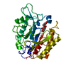
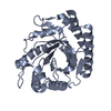
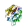
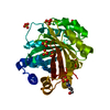
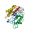
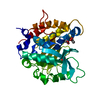
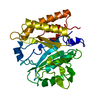
| Title | Crystal Structure of Cel5A (EG2) from Hypocrea jecorina (Trichoderma reesei) | ||||||
 Components Components | Endoglucanase EG-II | ||||||
 Keywords Keywords | HYDROLASE / Tim Barrel / Endoglucanase | ||||||
| Function / homology |  Function and homology information Function and homology informationcellulose binding / cellulase / cellulase activity / cellulose catabolic process / extracellular region Similarity search - Function | ||||||
| Biological species |  Hypocrea jecorina (fungus) Hypocrea jecorina (fungus) | ||||||
| Method |  X-RAY DIFFRACTION / SYNCHROTRON / Resolution: 2.05 Å X-RAY DIFFRACTION / SYNCHROTRON / Resolution: 2.05 Å | ||||||
 Authors Authors | Lee, T.M. / Farrow, M.F. / Kaiser, J.T. / Arnold, F.H. / Mayo, S.L. | ||||||
 Citation Citation | Journal: Protein Sci. / Year: 2011 Title: A structural study of Hypocrea jecorina Cel5A. Authors: Lee, T.M. / Farrow, M.F. / Arnold, F.H. / Mayo, S.L. | ||||||
| History |
|
- Structure visualization
| Structure viewer | Molecule: MolmilJmol/JSmol |
|---|
- Downloads & links
-Download
| PDBx/mmCIF format | 3qr3.cif.gz | 148.8 KB | Display | PDBx/mmCIF format |
|---|---|---|---|---|
| PDB format | pdb3qr3.ent.gz | 117.1 KB | Display | PDB format |
| PDBx/mmJSON format | 3qr3.json.gz | Tree view | PDBx/mmJSON format | |
| Others |  Other downloads Other downloads |
-Validation report
| Arichive directory | https://data.pdbj.org/pub/pdb/validation_reports/qr/3qr3ftp://data.pdbj.org/pub/pdb/validation_reports/qr/3qr3 | HTTPS FTP |
|---|
-Related structure data
| Similar structure data |
|---|


-Links
 PDBj
PDBj- Assembly
| Deposited unit | 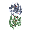
| ||||||||||||||||||||||||||||||||||||||||||||||||||||||||||||||
|---|---|---|---|---|---|---|---|---|---|---|---|---|---|---|---|---|---|---|---|---|---|---|---|---|---|---|---|---|---|---|---|---|---|---|---|---|---|---|---|---|---|---|---|---|---|---|---|---|---|---|---|---|---|---|---|---|---|---|---|---|---|---|---|
| 1 | 
| ||||||||||||||||||||||||||||||||||||||||||||||||||||||||||||||
| 2 | 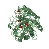
| ||||||||||||||||||||||||||||||||||||||||||||||||||||||||||||||
| Unit cell |
| ||||||||||||||||||||||||||||||||||||||||||||||||||||||||||||||
| Noncrystallographic symmetry (NCS) | NCS domain:
NCS domain segments:
NCS ensembles :
NCS oper:
| ||||||||||||||||||||||||||||||||||||||||||||||||||||||||||||||
| Details | BIOLOGICAL ASSEMBLY IS UNKNOWN AS PER AUTHORS |
-Components
| #1: Protein | Mass: 36589.031 Da / Num. of mol.: 2 / Fragment: CATALYTIC DOMAIN Source method: isolated from a genetically manipulated source Source: (gene. exp.) Hypocrea jecorina (fungus) / Gene: Cel5A, egl2 / Plasmid: pET22b / Production host:  #2: Chemical | ChemComp-SO4 /   Mass: 96.063 Da / Num. of mol.: 14 / Source method: obtained synthetically / Formula: SO4 Mass: 96.063 Da / Num. of mol.: 14 / Source method: obtained synthetically / Formula: SO4#3: Chemical | ChemComp-MG /   Mass: 24.305 Da / Num. of mol.: 4 / Source method: obtained synthetically / Formula: Mg Mass: 24.305 Da / Num. of mol.: 4 / Source method: obtained synthetically / Formula: Mg#4: Water | ChemComp-HOH / |  Mass: 18.015 Da / Num. of mol.: 503 / Source method: isolated from a natural source / Formula: H2O Mass: 18.015 Da / Num. of mol.: 503 / Source method: isolated from a natural source / Formula: H2OHas protein modification | Y | |
|---|
-Experimental details
-Experiment
| Experiment | Method: X-RAY DIFFRACTION / Number of used crystals: 1 |
|---|
- Sample preparation
| Crystal | Density Matthews: 2.16 Å3/Da / Density % sol: 42.95 % |
|---|---|
| Crystal grow | Temperature: 293 K / Method: vapor diffusion, sitting drop / pH: 5.6 Details: 0.1 M Sodium citrate, 1 M Magnesium sulfate, pH 5.6, VAPOR DIFFUSION, SITTING DROP, temperature 293K |
-Data collection
| Diffraction | Mean temperature: 293.15 K | |||||||||||||||||||||||||||||||||||||||||||||||||||||||||||||||||||||||||||||
|---|---|---|---|---|---|---|---|---|---|---|---|---|---|---|---|---|---|---|---|---|---|---|---|---|---|---|---|---|---|---|---|---|---|---|---|---|---|---|---|---|---|---|---|---|---|---|---|---|---|---|---|---|---|---|---|---|---|---|---|---|---|---|---|---|---|---|---|---|---|---|---|---|---|---|---|---|---|---|
| Diffraction source | Source: SYNCHROTRON / Site: SSRL  / Beamline: BL12-2 / Wavelength: 1.0332 Å / Beamline: BL12-2 / Wavelength: 1.0332 Å | |||||||||||||||||||||||||||||||||||||||||||||||||||||||||||||||||||||||||||||
| Detector | Type: DECTRIS PILATUS 6M / Detector: PIXEL / Date: Jan 14, 2011 / Details: Rh coated mirrors | |||||||||||||||||||||||||||||||||||||||||||||||||||||||||||||||||||||||||||||
| Radiation | Monochromator: Liquid nitrogen-cooled double crystal / Protocol: SINGLE WAVELENGTH / Monochromatic (M) / Laue (L): M / Scattering type: x-ray | |||||||||||||||||||||||||||||||||||||||||||||||||||||||||||||||||||||||||||||
| Radiation wavelength | Wavelength: 1.0332 Å / Relative weight: 1 | |||||||||||||||||||||||||||||||||||||||||||||||||||||||||||||||||||||||||||||
| Reflection | Resolution: 2.05→39.77 Å / Num. all: 40013 / Num. obs: 40013 / % possible obs: 98.8 % / Redundancy: 12.5 % / Rsym value: 0.081 / Net I/σ(I): 19.2 | |||||||||||||||||||||||||||||||||||||||||||||||||||||||||||||||||||||||||||||
| Reflection shell |
|
- Processing
| Software |
| |||||||||||||||||||||||||||||||||||||||||||||||||||||||||||||||||||||||||||||||||||||||||||||||||||||||||
|---|---|---|---|---|---|---|---|---|---|---|---|---|---|---|---|---|---|---|---|---|---|---|---|---|---|---|---|---|---|---|---|---|---|---|---|---|---|---|---|---|---|---|---|---|---|---|---|---|---|---|---|---|---|---|---|---|---|---|---|---|---|---|---|---|---|---|---|---|---|---|---|---|---|---|---|---|---|---|---|---|---|---|---|---|---|---|---|---|---|---|---|---|---|---|---|---|---|---|---|---|---|---|---|---|---|---|
| Refinement | Resolution: 2.05→39.767 Å / Cor.coef. Fo:Fc: 0.984 / Cor.coef. Fo:Fc free: 0.969 / SU ML: 0.24 / σ(F): 1.41 / Phase error: 26.7 / Stereochemistry target values: ML / Details: HYDROGENS HAVE BEEN ADDED IN THE RIDING POSITIONS
| |||||||||||||||||||||||||||||||||||||||||||||||||||||||||||||||||||||||||||||||||||||||||||||||||||||||||
| Solvent computation | Shrinkage radii: 0.9 Å / VDW probe radii: 1.11 Å / Solvent model: FLAT BULK SOLVENT MODEL / Bsol: 44.326 Å2 / ksol: 0.368 e/Å3 | |||||||||||||||||||||||||||||||||||||||||||||||||||||||||||||||||||||||||||||||||||||||||||||||||||||||||
| Displacement parameters |
| |||||||||||||||||||||||||||||||||||||||||||||||||||||||||||||||||||||||||||||||||||||||||||||||||||||||||
| Refinement step | Cycle: LAST / Resolution: 2.05→39.767 Å
| |||||||||||||||||||||||||||||||||||||||||||||||||||||||||||||||||||||||||||||||||||||||||||||||||||||||||
| Refine LS restraints |
| |||||||||||||||||||||||||||||||||||||||||||||||||||||||||||||||||||||||||||||||||||||||||||||||||||||||||
| Refine LS restraints NCS |
| |||||||||||||||||||||||||||||||||||||||||||||||||||||||||||||||||||||||||||||||||||||||||||||||||||||||||
| LS refinement shell |
|
