 Movie
Movie Controller


+ Open data

- Basic information
| Entry | Database: PDB / ID: 5d8f | ||||||
|---|---|---|---|---|---|---|---|
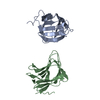
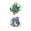
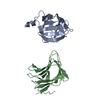
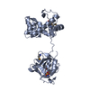
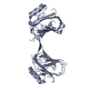
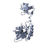
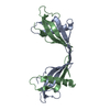
| Title | crystal structure of SSB and ssDNA complex from homo sapiens | ||||||
 Components Components |
| ||||||
 Keywords Keywords | DNA BINDING PROTEIN/DNA / single-strand DNA binding / DNA BINDING PROTEIN-DNA complex | ||||||
| Function / homology |  Function and homology information Function and homology informationSOSS complex / establishment of protein localization to telomere / positive regulation of telomere capping / G-rich strand telomeric DNA binding / mitotic G2/M transition checkpoint / response to ionizing radiation / RNA polymerase II transcribes snRNA genes / DNA polymerase binding / double-strand break repair via homologous recombination / site of double-strand break ...SOSS complex / establishment of protein localization to telomere / positive regulation of telomere capping / G-rich strand telomeric DNA binding / mitotic G2/M transition checkpoint / response to ionizing radiation / RNA polymerase II transcribes snRNA genes / DNA polymerase binding / double-strand break repair via homologous recombination / site of double-strand break / single-stranded DNA binding / chromosome, telomeric region / DNA repair / DNA damage response / DNA binding / nucleoplasm / nucleus / cytosol Similarity search - Function | ||||||
| Biological species |  Homo sapiens (human) Homo sapiens (human)synthetic construct (others) | ||||||
| Method |  X-RAY DIFFRACTION / SYNCHROTRON / MOLECULAR REPLACEMENT / Resolution: 2.35 Å X-RAY DIFFRACTION / SYNCHROTRON / MOLECULAR REPLACEMENT / Resolution: 2.35 Å | ||||||
 Authors Authors | Li, Y.H. / Gao, Z.Q. / Dong, Y.H. | ||||||
 Citation Citation | Journal: To Be Published Title: crystal structure of SSB and ssDNA complex from homo sapiens Authors: Li, Y.H. / Gao, Z.Q. / Dong, Y.H. | ||||||
| History |
|
- Structure visualization
| Structure viewer | Molecule: MolmilJmol/JSmol |
|---|
- Downloads & links
-Download
| PDBx/mmCIF format | 5d8f.cif.gz | 58.1 KB | Display | PDBx/mmCIF format |
|---|---|---|---|---|
| PDB format | pdb5d8f.ent.gz | 41.4 KB | Display | PDB format |
| PDBx/mmJSON format | 5d8f.json.gz | Tree view | PDBx/mmJSON format | |
| Others |  Other downloads Other downloads |
-Validation report
| Summary document | 5d8f_validation.pdf.gz | 454.5 KB | Display | wwPDB validaton report |
|---|---|---|---|---|
| Full document | 5d8f_full_validation.pdf.gz | 454.7 KB | Display | |
| Data in XML | 5d8f_validation.xml.gz | 9.8 KB | Display | |
| Data in CIF | 5d8f_validation.cif.gz | 12.5 KB | Display | |
| Arichive directory | https://data.pdbj.org/pub/pdb/validation_reports/d8/5d8fftp://data.pdbj.org/pub/pdb/validation_reports/d8/5d8f | HTTPS FTP |
-Related structure data
| Related structure data |  5d8eS S: Starting model for refinement |
|---|---|
| Similar structure data |

-Links
 PDBj
PDBj


- Assembly
| Deposited unit | 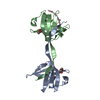
| ||||||||
|---|---|---|---|---|---|---|---|---|---|
| 1 |
| ||||||||
| Unit cell |
|
-Components
| #1: Protein | Mass: 12860.797 Da / Num. of mol.: 2 / Fragment: UNP residues 1-109 Source method: isolated from a genetically manipulated source Source: (gene. exp.) Homo sapiens (human) / Gene: NABP2, OBFC2B, SSB1, LP3587 / Plasmid: pET21a / Production host:  #2: DNA chain | | Mass: 2996.971 Da / Num. of mol.: 1 / Source method: obtained synthetically / Source: (synth.) synthetic construct (others) #3: Chemical |   Mass: 106.120 Da / Num. of mol.: 2 / Source method: obtained synthetically / Formula: C4H10O3 Mass: 106.120 Da / Num. of mol.: 2 / Source method: obtained synthetically / Formula: C4H10O3#4: Water | ChemComp-HOH / |  Mass: 18.015 Da / Num. of mol.: 14 / Source method: isolated from a natural source / Formula: H2O Mass: 18.015 Da / Num. of mol.: 14 / Source method: isolated from a natural source / Formula: H2O |
|---|
-Experimental details
-Experiment
| Experiment | Method: X-RAY DIFFRACTION / Number of used crystals: 1 |
|---|
- Sample preparation
| Crystal | Density Matthews: 2.93 Å3/Da / Density % sol: 57.96 % |
|---|---|
| Crystal grow | Temperature: 293 K / Method: vapor diffusion, sitting drop / pH: 6 / Details: 0.1M MES pH 6.0, 16% Jeffamine M-600 |
-Data collection
| Diffraction | Mean temperature: 100 K |
|---|---|
| Diffraction source | Source: SYNCHROTRON / Site: SSRF  / Beamline: BL17U / Wavelength: 0.9792 Å / Beamline: BL17U / Wavelength: 0.9792 Å |
| Detector | Type: ADSC QUANTUM 315r / Detector: CCD / Date: Nov 5, 2012 |
| Radiation | Protocol: SINGLE WAVELENGTH / Monochromatic (M) / Laue (L): M / Scattering type: x-ray |
| Radiation wavelength | Wavelength: 0.9792 Å / Relative weight: 1 |
| Reflection | Resolution: 2.35→50 Å / Num. all: 14419 / Num. obs: 14419 / % possible obs: 99.3 % / Redundancy: 19.9 % / Rmerge(I) obs: 0.063 / Net I/σ(I): 73 |
| Reflection shell | Resolution: 2.35→2.39 Å / Redundancy: 21.5 % / Rmerge(I) obs: 0.507 / Mean I/σ(I) obs: 9.16 / % possible all: 100 |
- Processing
| Software |
| ||||||||||||||||||||||||||||||||||||||||||
|---|---|---|---|---|---|---|---|---|---|---|---|---|---|---|---|---|---|---|---|---|---|---|---|---|---|---|---|---|---|---|---|---|---|---|---|---|---|---|---|---|---|---|---|
| Refinement | Method to determine structure: MOLECULAR REPLACEMENT Starting model: 5D8E Resolution: 2.35→41.91 Å / SU ML: 0.3 / Cross valid method: FREE R-VALUE / σ(F): 1.35 / Phase error: 30.81 / Stereochemistry target values: ML
| ||||||||||||||||||||||||||||||||||||||||||
| Solvent computation | Shrinkage radii: 0.9 Å / VDW probe radii: 1.11 Å / Solvent model: FLAT BULK SOLVENT MODEL | ||||||||||||||||||||||||||||||||||||||||||
| Refinement step | Cycle: LAST / Resolution: 2.35→41.91 Å
| ||||||||||||||||||||||||||||||||||||||||||
| Refine LS restraints |
| ||||||||||||||||||||||||||||||||||||||||||
| LS refinement shell |
|
