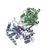
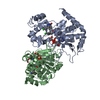
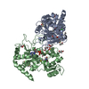
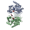
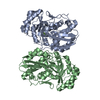
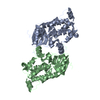
Entry Database : PDB / ID : 5d7qTitle Crystal structure of human Sirt2 in complex with ADPR and CHIC35 NAD-dependent protein deacetylase sirtuin-2 Keywords / / / Function / homology Function Domain/homology Component
/ / / / / / / / / / / / / / / / / / / / / / / / / / / / / / / / / / / / / / / / / / / / / / / / / / / / / / / / / / / / / / / / / / / / / / / / / / / / / / / / / / / / / / / / / / / / / / / / / / / / / / / / / / / / / / / / / / / / / / / / / / / / Biological species Homo sapiens (human)Method / / Resolution : 2.01 Å Authors Rumpf, T. / Gerhardt, S. / Einsle, O. / Jung, M. Funding support Organization Grant number Country German Research Foundation Ju295/8-1 German Research Foundation SFB992 Medical Epigenetics, Project Z02
Journal : Acta Crystallogr.,Sect.F / Year : 2015Title : Seeding for sirtuins: microseed matrix seeding to obtain crystals of human Sirt3 and Sirt2 suitable for soaking.Authors : Rumpf, T. / Gerhardt, S. / Einsle, O. / Jung, M. History Deposition Aug 14, 2015 Deposition site / Processing site Revision 1.0 Dec 2, 2015 Provider / Type Revision 1.1 Dec 9, 2015 Group Revision 1.2 Jan 10, 2024 Group Author supporting evidence / Data collection ... Author supporting evidence / Data collection / Database references / Refinement description / Structure summary Category chem_comp / chem_comp_atom ... chem_comp / chem_comp_atom / chem_comp_bond / database_2 / pdbx_audit_support / pdbx_initial_refinement_model Item _chem_comp.pdbx_synonyms / _database_2.pdbx_DOI ... _chem_comp.pdbx_synonyms / _database_2.pdbx_DOI / _database_2.pdbx_database_accession / _pdbx_audit_support.funding_organization
Show all Show less
 Movie
Movie Controller


 Open data
 Basic information
 Components
 Keywords
 Function and homology information
 Homo sapiens (human)
 X-RAY DIFFRACTION /
  Authors
 Germany, 2items
  Citation
 Structure visualization
 Downloads & links
 Other downloads
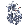


 PDBj
PDBj


 Assembly
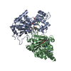


 Mass: 65.409 Da / Num. of mol.: 2 / Source method: obtained synthetically / Formula: Zn
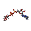
 Mass: 559.316 Da / Num. of mol.: 2 / Source method: obtained synthetically / Formula: C15H23N5O14P2
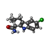
 Mass: 262.735 Da / Num. of mol.: 4 / Source method: obtained synthetically / Formula: C14H15ClN2O
 Mass: 18.015 Da / Num. of mol.: 137 / Source method: isolated from a natural source / Formula: H2O
 Sample preparation
 Processing