 Movie
Movie Controller

[English] 日本語
 Yorodumi
- PDB-3fyy: Crystal structure of divergent enolase from Oceanobacillus iheyen... -
+ Open data

- Basic information
| Entry | Database: PDB / ID: 3fyy | ||||||
|---|---|---|---|---|---|---|---|
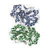
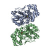
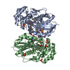
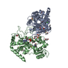
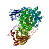
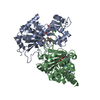
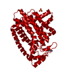
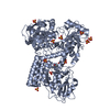
| Title | Crystal structure of divergent enolase from Oceanobacillus iheyensis complexed with Mg | ||||||
 Components Components | Muconate cycloisomerase | ||||||
 Keywords Keywords | ISOMERASE / divergent enolase / NYSGXRC / target 9375a / Structural Genomics / PSI-2 / Protein Structure Initiative / New York SGX Research Center for Structural Genomics | ||||||
| Function / homology |  Function and homology information Function and homology informationgalactarate dehydratase (D-threo-forming) / lyase activity / metal ion binding Similarity search - Function | ||||||
| Biological species |  Oceanobacillus iheyensis HTE831 (bacteria) Oceanobacillus iheyensis HTE831 (bacteria) | ||||||
| Method |  X-RAY DIFFRACTION / SYNCHROTRON / MOLECULAR REPLACEMENT / Resolution: 1.8 Å X-RAY DIFFRACTION / SYNCHROTRON / MOLECULAR REPLACEMENT / Resolution: 1.8 Å | ||||||
 Authors Authors | Fedorov, A.A. / Fedorov, E.V. / Sauder, J.M. / Burley, S.K. / Gerlt, J.A. / Almo, S.C. / New York SGX Research Center for Structural Genomics (NYSGXRC) | ||||||
 Citation Citation | Journal: Biochemistry / Year: 2009 Title: Computation-facilitated assignment of the function in the enolase superfamily: a regiochemically distinct galactarate dehydratase from Oceanobacillus iheyensis . Authors: Rakus, J.F. / Kalyanaraman, C. / Fedorov, A.A. / Fedorov, E.V. / Mills-Groninger, F.P. / Toro, R. / Bonanno, J. / Bain, K. / Sauder, J.M. / Burley, S.K. / Almo, S.C. / Jacobson, M.P. / Gerlt, J.A. | ||||||
| History |
|
- Structure visualization
| Structure viewer | Molecule: MolmilJmol/JSmol |
|---|
- Downloads & links
-Download
| PDBx/mmCIF format | 3fyy.cif.gz | 172.4 KB | Display | PDBx/mmCIF format |
|---|---|---|---|---|
| PDB format | pdb3fyy.ent.gz | 135.6 KB | Display | PDB format |
| PDBx/mmJSON format | 3fyy.json.gz | Tree view | PDBx/mmJSON format | |
| Others |  Other downloads Other downloads |
-Validation report
| Arichive directory | https://data.pdbj.org/pub/pdb/validation_reports/fy/3fyyftp://data.pdbj.org/pub/pdb/validation_reports/fy/3fyy | HTTPS FTP |
|---|
-Related structure data
| Related structure data |  2oqySC  3es7C  3es8C  3hpfC S: Starting model for refinement C: citing same article ( |
|---|---|
| Similar structure data | |
| Other databases |


-Links
 PDBj
PDBj
- Assembly
| Deposited unit | 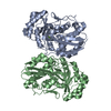
| ||||||||
|---|---|---|---|---|---|---|---|---|---|
| 1 |
| ||||||||
| 2 | 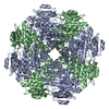
| ||||||||
| Unit cell |
| ||||||||
| Details | THE BIOLOGICAL ASSEMBLY IS A DIMER |
-Components
| #1: Protein | Mass: 44403.367 Da / Num. of mol.: 2 Source method: isolated from a genetically manipulated source Source: (gene. exp.) Oceanobacillus iheyensis HTE831 (bacteria)Strain: DSM 14371, JCM 11309, KCTC 3954, HTE831 / Gene: OB2843 / Production host: #2: Chemical | ChemComp-MG /   Mass: 24.305 Da / Num. of mol.: 4 / Source method: obtained synthetically / Formula: Mg Mass: 24.305 Da / Num. of mol.: 4 / Source method: obtained synthetically / Formula: Mg#3: Water | ChemComp-HOH / |  Mass: 18.015 Da / Num. of mol.: 578 / Source method: isolated from a natural source / Formula: H2O Mass: 18.015 Da / Num. of mol.: 578 / Source method: isolated from a natural source / Formula: H2O |
|---|
-Experimental details
-Experiment
| Experiment | Method: X-RAY DIFFRACTION / Number of used crystals: 1 |
|---|
- Sample preparation
| Crystal | Density Matthews: 2.5 Å3/Da / Density % sol: 50.75 % |
|---|---|
| Crystal grow | Temperature: 293 K / Method: vapor diffusion, hanging drop / pH: 7 Details: 1.0 M K/Na tartrate, 0.1M Tris, 0.2M Lithium sulfate, pH 7.0, VAPOR DIFFUSION, HANGING DROP, temperature 293.0K |
-Data collection
| Diffraction | Mean temperature: 100 K |
|---|---|
| Diffraction source | Source: SYNCHROTRON / Site: NSLS  / Beamline: X4A / Wavelength: 0.97915 Å / Beamline: X4A / Wavelength: 0.97915 Å |
| Detector | Type: ADSC QUANTUM 4 / Detector: CCD / Date: Nov 23, 2008 |
| Radiation | Monochromator: Si(111) CHANNEL / Protocol: SINGLE WAVELENGTH / Monochromatic (M) / Laue (L): M / Scattering type: x-ray |
| Radiation wavelength | Wavelength: 0.97915 Å / Relative weight: 1 |
| Reflection | Resolution: 1.8→25 Å / Num. all: 79723 / Num. obs: 79723 / % possible obs: 98.9 % / Observed criterion σ(F): 0 / Observed criterion σ(I): 0 / Biso Wilson estimate: 31.5 Å2 / Rmerge(I) obs: 0.065 |
- Processing
| Software |
| ||||||||||||||||||||||||||||||||||||
|---|---|---|---|---|---|---|---|---|---|---|---|---|---|---|---|---|---|---|---|---|---|---|---|---|---|---|---|---|---|---|---|---|---|---|---|---|---|
| Refinement | Method to determine structure: MOLECULAR REPLACEMENT Starting model: PDB entry 2OQY Resolution: 1.8→24.72 Å / Rfactor Rfree error: 0.004 / Data cutoff high absF: 2084274.97 / Data cutoff low absF: 0 / Isotropic thermal model: RESTRAINED / Cross valid method: THROUGHOUT / σ(F): 0 / σ(I): 0 / Stereochemistry target values: Engh & Huber
| ||||||||||||||||||||||||||||||||||||
| Solvent computation | Solvent model: FLAT MODEL / Bsol: 59.1595 Å2 / ksol: 0.362459 e/Å3 | ||||||||||||||||||||||||||||||||||||
| Displacement parameters | Biso mean: 35.5 Å2
| ||||||||||||||||||||||||||||||||||||
| Refine analyze |
| ||||||||||||||||||||||||||||||||||||
| Refinement step | Cycle: LAST / Resolution: 1.8→24.72 Å
| ||||||||||||||||||||||||||||||||||||
| Refine LS restraints |
| ||||||||||||||||||||||||||||||||||||
| LS refinement shell | Resolution: 1.8→1.86 Å / Rfactor Rfree error: 0.017 / Total num. of bins used: 10
| ||||||||||||||||||||||||||||||||||||
| Xplor file |
|
