Resolution: 2.7→2.75 Å / Redundancy: 11.2 % / Rmerge(I) obs: 0.621 / Mean I/σ(I) obs: 5 / % possible all: 100
-
Processing
Software
Name
Version
Classification
REFMAC
5.8.0073
refinement
BLU-MAX
datacollection
HKL-3000
phasing
HKL-3000
datascaling
PHENIX
phasing
Refinement
Method to determine structure: SAD / Resolution: 2.7→28.55 Å / Cor.coef. Fo:Fc: 0.944 / Cor.coef. Fo:Fc free: 0.908 / SU B: 28.361 / SU ML: 0.283 / Cross valid method: THROUGHOUT / ESU R Free: 0.407 / Stereochemistry target values: MAXIMUM LIKELIHOOD / Details: HYDROGENS HAVE BEEN ADDED IN THE RIDING POSITIONS
Rfactor
Num. reflection
% reflection
Selection details
Rfree
0.25555
1535
10.1 %
RANDOM
Rwork
0.1941
-
-
-
obs
0.19991
13687
99.8 %
-
Solvent computation
Ion probe radii: 0.8 Å / Shrinkage radii: 0.8 Å / VDW probe radii: 1.2 Å / Solvent model: MASK
 Movie
Movie Controller

 Yorodumi
 Open data
 Basic information
 Components
 Keywords
 Function and homology information
 Vibrio vulnificus (bacteria)
 X-RAY DIFFRACTION /
  Authors
 Citation
 Structure visualization
 Downloads & links
 Other downloads


 PDBj
PDBj


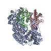
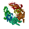
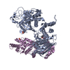
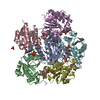
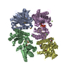
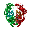
 Assembly
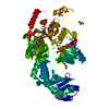

 Mass: 22.990 Da / Num. of mol.: 1 / Source method: obtained synthetically / Formula: Na
 Mass: 506.196 Da / Num. of mol.: 1 / Source method: obtained synthetically / Formula: C10H17N6O12P3 / Comment: AMP-PNP, energy-carrying molecule analogue*YM
 Mass: 18.015 Da / Num. of mol.: 84 / Source method: isolated from a natural source / Formula: H2O
 Sample preparation
 / Beamline: 21-ID-G / Wavelength: 0.97856 Å
 Processing