 Movie
Movie Controller

[English] 日本語
 Yorodumi
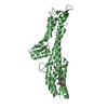
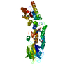
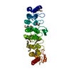
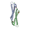
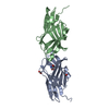
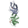
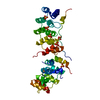
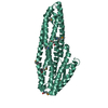
- PDB-5d55: Crystal structure of the E. coli Hda pilus minor tip subunit, HdaB -
+ Open data

- Basic information
| Entry | Database: PDB / ID: 5d55 | ||||||
|---|---|---|---|---|---|---|---|
| Title | Crystal structure of the E. coli Hda pilus minor tip subunit, HdaB | ||||||
 Components Components | HdaB,HdaA (Adhesin), HUS-associated diffuse adherence | ||||||
 Keywords Keywords | CELL ADHESION / Biofilm / chaperone-usher / Hda / pilus | ||||||
| Function / homology |  Function and homology information Function and homology informationEnterobacteria AfaD invasin / Enterobacteria AfaD invasin protein / Dr adhesin / Dr-adhesin superfamily / Adhesion domain superfamily / Immunoglobulin-like / Sandwich / Mainly Beta Similarity search - Domain/homology | ||||||
| Biological species |  | ||||||
| Method |  X-RAY DIFFRACTION / SYNCHROTRON / MOLECULAR REPLACEMENT / Resolution: 2 Å X-RAY DIFFRACTION / SYNCHROTRON / MOLECULAR REPLACEMENT / Resolution: 2 Å | ||||||
 Authors Authors | Lee, W.-C. / Garnett, J.A. / Matthews, S.J. | ||||||
 Citation Citation | Journal: Protein Sci. / Year: 2016 Title: Crystal structure and analysis of HdaB: The enteroaggregative Escherichia coli AAF/IV pilus tip protein. Authors: Lee, W.C. / Matthews, S. / Garnett, J.A. | ||||||
| History |
|
- Structure visualization
| Structure viewer | Molecule: MolmilJmol/JSmol |
|---|
- Downloads & links
-Download
| PDBx/mmCIF format | 5d55.cif.gz | 130.8 KB | Display | PDBx/mmCIF format |
|---|---|---|---|---|
| PDB format | pdb5d55.ent.gz | 101.5 KB | Display | PDB format |
| PDBx/mmJSON format | 5d55.json.gz | Tree view | PDBx/mmJSON format | |
| Others |  Other downloads Other downloads |
-Validation report
| Summary document | 5d55_validation.pdf.gz | 461.3 KB | Display | wwPDB validaton report |
|---|---|---|---|---|
| Full document | 5d55_full_validation.pdf.gz | 470.2 KB | Display | |
| Data in XML | 5d55_validation.xml.gz | 17.6 KB | Display | |
| Data in CIF | 5d55_validation.cif.gz | 24.6 KB | Display | |
| Arichive directory | https://data.pdbj.org/pub/pdb/validation_reports/d5/5d55ftp://data.pdbj.org/pub/pdb/validation_reports/d5/5d55 | HTTPS FTP |
-Related structure data
| Related structure data | 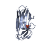 4or1S S: Starting model for refinement |
|---|---|
| Similar structure data |
-Links
 PDBj
PDBj


- Assembly
| Deposited unit | 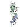
| ||||||||
|---|---|---|---|---|---|---|---|---|---|
| 1 |
| ||||||||
| Unit cell |
|
-Components
| #1: Protein | Mass: 17488.668 Da / Num. of mol.: 2 Fragment: UNP residues 24-142,UNP residues 18-34,UNP residues 24-142,UNP residues 18-34 Source method: isolated from a genetically manipulated source Source: (gene. exp.) #2: Chemical |   Mass: 189.100 Da / Num. of mol.: 3 / Source method: obtained synthetically / Formula: C6H5O7 Mass: 189.100 Da / Num. of mol.: 3 / Source method: obtained synthetically / Formula: C6H5O7#3: Chemical | ChemComp-IOD / |   Mass: 126.904 Da / Num. of mol.: 1 / Source method: obtained synthetically / Formula: I Mass: 126.904 Da / Num. of mol.: 1 / Source method: obtained synthetically / Formula: I#4: Water | ChemComp-HOH / |  Mass: 18.015 Da / Num. of mol.: 229 / Source method: isolated from a natural source / Formula: H2O Mass: 18.015 Da / Num. of mol.: 229 / Source method: isolated from a natural source / Formula: H2OHas protein modification | Y | |
|---|
-Experimental details
-Experiment
| Experiment | Method: X-RAY DIFFRACTION |
|---|
- Sample preparation
| Crystal | Density Matthews: 2.8 Å3/Da / Density % sol: 56.15 % |
|---|---|
| Crystal grow | Temperature: 293.15 K / Method: vapor diffusion, sitting drop / pH: 5 / Details: 0.2M Ammonium Citrate, 20% PEG 3350 |
-Data collection
| Diffraction | Mean temperature: 100 K |
|---|---|
| Diffraction source | Source: SYNCHROTRON / Site: Diamond  / Beamline: I24 / Wavelength: 1.6531 Å / Beamline: I24 / Wavelength: 1.6531 Å |
| Detector | Type: DECTRIS PILATUS 6M / Detector: PIXEL / Date: Aug 8, 2012 |
| Radiation | Protocol: SINGLE WAVELENGTH / Monochromatic (M) / Laue (L): M / Scattering type: x-ray |
| Radiation wavelength | Wavelength: 1.6531 Å / Relative weight: 1 |
| Reflection | Resolution: 2→79.4 Å / Num. obs: 26601 / % possible obs: 98.1 % / Redundancy: 3.6 % / Biso Wilson estimate: 31.636 Å2 / Rmerge(I) obs: 0.08 / Net I/σ(I): 9.3 |
| Reflection shell | Resolution: 2→2.11 Å / Redundancy: 3.1 % / Rmerge(I) obs: 0.371 / Mean I/σ(I) obs: 2.7 / % possible all: 95.1 |
- Processing
| Software |
| ||||||||||||||||||||||||||||||||||||||||||||||||||||||||||||||||||||||||||||||||||||||||||||||||||||||||||||||||||||||||||||||||||||||||||||||||||||||||||||||||||||||||||||||||||||||
|---|---|---|---|---|---|---|---|---|---|---|---|---|---|---|---|---|---|---|---|---|---|---|---|---|---|---|---|---|---|---|---|---|---|---|---|---|---|---|---|---|---|---|---|---|---|---|---|---|---|---|---|---|---|---|---|---|---|---|---|---|---|---|---|---|---|---|---|---|---|---|---|---|---|---|---|---|---|---|---|---|---|---|---|---|---|---|---|---|---|---|---|---|---|---|---|---|---|---|---|---|---|---|---|---|---|---|---|---|---|---|---|---|---|---|---|---|---|---|---|---|---|---|---|---|---|---|---|---|---|---|---|---|---|---|---|---|---|---|---|---|---|---|---|---|---|---|---|---|---|---|---|---|---|---|---|---|---|---|---|---|---|---|---|---|---|---|---|---|---|---|---|---|---|---|---|---|---|---|---|---|---|---|---|
| Refinement | Method to determine structure: MOLECULAR REPLACEMENT Starting model: 4OR1 Resolution: 2→79.4 Å / Cor.coef. Fo:Fc: 0.952 / Cor.coef. Fo:Fc free: 0.927 / SU B: 7.378 / SU ML: 0.108 / Cross valid method: THROUGHOUT / ESU R: 0.181 / ESU R Free: 0.169 / Stereochemistry target values: MAXIMUM LIKELIHOOD / Details: HYDROGENS HAVE BEEN USED IF PRESENT IN THE INPUT
| ||||||||||||||||||||||||||||||||||||||||||||||||||||||||||||||||||||||||||||||||||||||||||||||||||||||||||||||||||||||||||||||||||||||||||||||||||||||||||||||||||||||||||||||||||||||
| Solvent computation | Ion probe radii: 0.8 Å / Shrinkage radii: 0.8 Å / VDW probe radii: 1.2 Å / Solvent model: MASK | ||||||||||||||||||||||||||||||||||||||||||||||||||||||||||||||||||||||||||||||||||||||||||||||||||||||||||||||||||||||||||||||||||||||||||||||||||||||||||||||||||||||||||||||||||||||
| Displacement parameters | Biso mean: 34.393 Å2
| ||||||||||||||||||||||||||||||||||||||||||||||||||||||||||||||||||||||||||||||||||||||||||||||||||||||||||||||||||||||||||||||||||||||||||||||||||||||||||||||||||||||||||||||||||||||
| Refinement step | Cycle: 1 / Resolution: 2→79.4 Å
| ||||||||||||||||||||||||||||||||||||||||||||||||||||||||||||||||||||||||||||||||||||||||||||||||||||||||||||||||||||||||||||||||||||||||||||||||||||||||||||||||||||||||||||||||||||||
| Refine LS restraints |
|