 Movie
Movie Controller

[English] 日本語
 Yorodumi
- PDB-4or1: Structure and mechanism of fibronectin binding and biofilm format... -
+ Open data

- Basic information
| Entry | Database: PDB / ID: 4or1 | ||||||
|---|---|---|---|---|---|---|---|
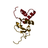
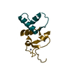
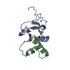
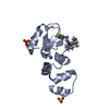
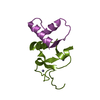
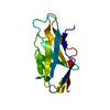
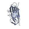
| Title | Structure and mechanism of fibronectin binding and biofilm formation of enteroaggregative Escherischia coli AAF fimbriae | ||||||
 Components Components | Invasin homolog AafB, Major fimbrial subunit of aggregative adherence fimbria II AafA chimeric construct | ||||||
 Keywords Keywords | CELL ADHESION / biofilm / chaperone-usher / pilus / adhesion / fibronectin / fimbriae / fibre / immunoglobulin-like / Outer membrane pilus | ||||||
| Function / homology |  Function and homology information Function and homology informationEnterobacteria AfaD invasin / Enterobacteria AfaD invasin protein / Dr adhesin / Dr-adhesin superfamily / Adhesion domain superfamily / Immunoglobulin-like / Sandwich / Mainly Beta Similarity search - Domain/homology | ||||||
| Biological species |  | ||||||
| Method |  X-RAY DIFFRACTION / SYNCHROTRON / MOLECULAR REPLACEMENT / Resolution: 3 Å X-RAY DIFFRACTION / SYNCHROTRON / MOLECULAR REPLACEMENT / Resolution: 3 Å | ||||||
 Authors Authors | Lee, W.-C. / Garnett, J.A. / Yang, Y. / Matthews, S. | ||||||
 Citation Citation | Journal: Plos Pathog. / Year: 2014 Title: Structural insight into host recognition by aggregative adherence fimbriae of enteroaggregative Escherichia coli. Authors: Berry, A.A. / Yang, Y. / Pakharukova, N. / Garnett, J.A. / Lee, W.C. / Cota, E. / Marchant, J. / Roy, S. / Tuittila, M. / Liu, B. / Inman, K.G. / Ruiz-Perez, F. / Mandomando, I. / Nataro, J. ...Authors: Berry, A.A. / Yang, Y. / Pakharukova, N. / Garnett, J.A. / Lee, W.C. / Cota, E. / Marchant, J. / Roy, S. / Tuittila, M. / Liu, B. / Inman, K.G. / Ruiz-Perez, F. / Mandomando, I. / Nataro, J.P. / Zavialov, A.V. / Matthews, S. | ||||||
| History |
|
- Structure visualization
| Structure viewer | Molecule: MolmilJmol/JSmol |
|---|
- Downloads & links
-Download
| PDBx/mmCIF format | 4or1.cif.gz | 62.6 KB | Display | PDBx/mmCIF format |
|---|---|---|---|---|
| PDB format | pdb4or1.ent.gz | 45.6 KB | Display | PDB format |
| PDBx/mmJSON format | 4or1.json.gz | Tree view | PDBx/mmJSON format | |
| Others |  Other downloads Other downloads |
-Validation report
| Arichive directory | https://data.pdbj.org/pub/pdb/validation_reports/or/4or1ftp://data.pdbj.org/pub/pdb/validation_reports/or/4or1 | HTTPS FTP |
|---|
-Related structure data
| Related structure data | 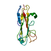 2mpvC 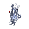 4ph8C  4phxC  2axwS S: Starting model for refinement C: citing same article ( |
|---|---|
| Similar structure data |


-Links
 PDBj
PDBj- Assembly
| Deposited unit | 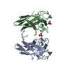
| ||||||||
|---|---|---|---|---|---|---|---|---|---|
| 1 | 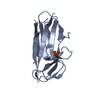
| ||||||||
| 2 | 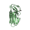
| ||||||||
| Unit cell |
|
-Components
| #1: Protein | Mass: 17186.150 Da / Num. of mol.: 2 Fragment: AAF/II pilus minor pilin, unp residues 24-146 and unp residues 25-34 Source method: isolated from a genetically manipulated source Source: (gene. exp.) #2: Chemical | ChemComp-SO4 / |   Mass: 96.063 Da / Num. of mol.: 1 / Source method: obtained synthetically / Formula: SO4 Mass: 96.063 Da / Num. of mol.: 1 / Source method: obtained synthetically / Formula: SO4#3: Chemical | ChemComp-ACT / |   Mass: 59.044 Da / Num. of mol.: 1 / Source method: obtained synthetically / Formula: C2H3O2 Mass: 59.044 Da / Num. of mol.: 1 / Source method: obtained synthetically / Formula: C2H3O2#4: Water | ChemComp-HOH / |  Mass: 18.015 Da / Num. of mol.: 2 / Source method: isolated from a natural source / Formula: H2O Mass: 18.015 Da / Num. of mol.: 2 / Source method: isolated from a natural source / Formula: H2OHas protein modification | Y | |
|---|
-Experimental details
-Experiment
| Experiment | Method: X-RAY DIFFRACTION / Number of used crystals: 1 |
|---|
- Sample preparation
| Crystal | Density Matthews: 3.13 Å3/Da / Density % sol: 60.64 % |
|---|---|
| Crystal grow | Temperature: 293 K / Method: vapor diffusion, sitting drop / pH: 5.5 Details: 0.1 M BIS-TRIS, 0.2 M LiSO4, 25% PEG3350., pH 5.5, VAPOR DIFFUSION, SITTING DROP, temperature 293K |
-Data collection
| Diffraction | Mean temperature: 100 K |
|---|---|
| Diffraction source | Source: SYNCHROTRON / Site: Diamond  / Beamline: I24 / Wavelength: 0.9778 Å / Beamline: I24 / Wavelength: 0.9778 Å |
| Detector | Type: PSI PILATUS 6M / Detector: PIXEL / Date: Oct 2, 2011 |
| Radiation | Monochromator: double crystal / Protocol: SINGLE WAVELENGTH / Monochromatic (M) / Laue (L): M / Scattering type: x-ray |
| Radiation wavelength | Wavelength: 0.9778 Å / Relative weight: 1 |
| Reflection | Resolution: 3→51.24 Å / Num. all: 9081 / Num. obs: 9081 / % possible obs: 97 % / Observed criterion σ(F): 2 / Observed criterion σ(I): 2 / Redundancy: 2.9 % / Biso Wilson estimate: 74.4 Å2 / Rsym value: 0.111 / Net I/σ(I): 6.5 |
| Reflection shell | Resolution: 3→3.16 Å / Redundancy: 2.9 % / Mean I/σ(I) obs: 2.6 / Num. unique all: 1302 / Rsym value: 0.374 / % possible all: 98.2 |
- Processing
| Software |
| |||||||||||||||||||||||||||||||||||||||||||||
|---|---|---|---|---|---|---|---|---|---|---|---|---|---|---|---|---|---|---|---|---|---|---|---|---|---|---|---|---|---|---|---|---|---|---|---|---|---|---|---|---|---|---|---|---|---|---|
| Refinement | Method to determine structure: MOLECULAR REPLACEMENT Starting model: PDB ENTRY 2AXW Resolution: 3→51.24 Å / Cor.coef. Fo:Fc: 0.915 / Cor.coef. Fo:Fc free: 0.905 / SU B: 14.549 / SU ML: 0.26 / Cross valid method: THROUGHOUT / σ(F): 2 / ESU R: 0.995 / ESU R Free: 0.364 / Stereochemistry target values: MAXIMUM LIKELIHOOD / Details: HYDROGENS HAVE BEEN USED IF PRESENT IN THE INPUT
| |||||||||||||||||||||||||||||||||||||||||||||
| Solvent computation | Ion probe radii: 0.8 Å / Shrinkage radii: 0.8 Å / VDW probe radii: 1.2 Å / Solvent model: MASK | |||||||||||||||||||||||||||||||||||||||||||||
| Displacement parameters | Biso mean: 56.38 Å2
| |||||||||||||||||||||||||||||||||||||||||||||
| Refinement step | Cycle: LAST / Resolution: 3→51.24 Å
| |||||||||||||||||||||||||||||||||||||||||||||
| Refine LS restraints |
| |||||||||||||||||||||||||||||||||||||||||||||
| LS refinement shell | Resolution: 3→3.078 Å / Total num. of bins used: 20
|
