 Movie
Movie Controller

[English] 日本語
 Yorodumi
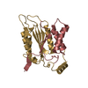
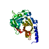
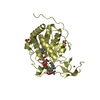
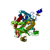
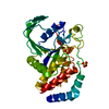
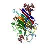
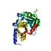
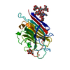
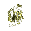
- PDB-5d1i: Structure of Cyclic nucleotide-binding-like protein from Brucella... -
+ Open data

- Basic information
| Entry | Database: PDB / ID: 5d1i | ||||||
|---|---|---|---|---|---|---|---|
| Title | Structure of Cyclic nucleotide-binding-like protein from Brucella abortus bv. 1 str. 9-941 | ||||||
 Components Components | Cyclic nucleotide-binding protein | ||||||
 Keywords Keywords | UNKNOWN FUNCTION / beta barrel | ||||||
| Function / homology |  Function and homology information Function and homology information | ||||||
| Biological species |  Brucella abortus biovar 1 (bacteria) Brucella abortus biovar 1 (bacteria) | ||||||
| Method |  X-RAY DIFFRACTION / SYNCHROTRON / SAD / Resolution: 2 Å X-RAY DIFFRACTION / SYNCHROTRON / SAD / Resolution: 2 Å | ||||||
 Authors Authors | He, Z. / Dong, J. / Li, X. / Gao, Y. | ||||||
 Citation Citation | Journal: Biochem.Biophys.Res.Commun. / Year: 2015 Title: Crystal structure of cyclic nucleotide-binding-like protein from Brucella abortus Authors: He, Z. / Gao, Y. / Dong, J. / Ke, Y. / Li, X. / Chen, Z. / Zhang, X.C. | ||||||
| History |
|
- Structure visualization
| Structure viewer | Molecule: MolmilJmol/JSmol |
|---|
- Downloads & links
-Download
| PDBx/mmCIF format | 5d1i.cif.gz | 110.3 KB | Display | PDBx/mmCIF format |
|---|---|---|---|---|
| PDB format | pdb5d1i.ent.gz | 85 KB | Display | PDB format |
| PDBx/mmJSON format | 5d1i.json.gz | Tree view | PDBx/mmJSON format | |
| Others |  Other downloads Other downloads |
-Validation report
| Arichive directory | https://data.pdbj.org/pub/pdb/validation_reports/d1/5d1iftp://data.pdbj.org/pub/pdb/validation_reports/d1/5d1i | HTTPS FTP |
|---|
-Related structure data
| Similar structure data |
|---|

-Links
 PDBj
PDBj


- Assembly
| Deposited unit | 
| |||||||||
|---|---|---|---|---|---|---|---|---|---|---|
| 1 |
| |||||||||
| Unit cell |
| |||||||||
| Components on special symmetry positions |
|
-Components
| #1: Protein | Mass: 14455.212 Da / Num. of mol.: 2 / Fragment: UNP residues 1-123 Source method: isolated from a genetically manipulated source Source: (gene. exp.) Brucella abortus biovar 1 (bacteria) / Strain: 9-941 / Gene: BruAb1_1980 / Plasmid: pGEX-6P-1 / Production host: #2: Water | ChemComp-HOH / |  Mass: 18.015 Da / Num. of mol.: 171 / Source method: isolated from a natural source / Formula: H2O Mass: 18.015 Da / Num. of mol.: 171 / Source method: isolated from a natural source / Formula: H2OHas protein modification | Y | |
|---|
-Experimental details
-Experiment
| Experiment | Method: X-RAY DIFFRACTION / Number of used crystals: 1 |
|---|
- Sample preparation
| Crystal | Density Matthews: 2.09 Å3/Da / Density % sol: 41.14 % |
|---|---|
| Crystal grow | Temperature: 293 K / Method: vapor diffusion, hanging drop / pH: 7.4 Details: Potassium sodium tartrate tetrahydrate,olyethylene glycol 3350, KCl |
-Data collection
| Diffraction | Mean temperature: 100 K | |||||||||||||||||||||||||||||||||||||||||||||||||||||||||||||||||||||||||||||||||||||||||||||||||||
|---|---|---|---|---|---|---|---|---|---|---|---|---|---|---|---|---|---|---|---|---|---|---|---|---|---|---|---|---|---|---|---|---|---|---|---|---|---|---|---|---|---|---|---|---|---|---|---|---|---|---|---|---|---|---|---|---|---|---|---|---|---|---|---|---|---|---|---|---|---|---|---|---|---|---|---|---|---|---|---|---|---|---|---|---|---|---|---|---|---|---|---|---|---|---|---|---|---|---|---|---|
| Diffraction source | Source: SYNCHROTRON / Site: SSRF  / Beamline: BL17U / Wavelength: 0.9762 Å / Beamline: BL17U / Wavelength: 0.9762 Å | |||||||||||||||||||||||||||||||||||||||||||||||||||||||||||||||||||||||||||||||||||||||||||||||||||
| Detector | Type: ADSC QUANTUM 315r / Detector: CCD / Date: Mar 27, 2015 / Details: mirrors | |||||||||||||||||||||||||||||||||||||||||||||||||||||||||||||||||||||||||||||||||||||||||||||||||||
| Radiation | Monochromator: double crystal / Protocol: SINGLE WAVELENGTH / Monochromatic (M) / Laue (L): M / Scattering type: x-ray | |||||||||||||||||||||||||||||||||||||||||||||||||||||||||||||||||||||||||||||||||||||||||||||||||||
| Radiation wavelength | Wavelength: 0.9762 Å / Relative weight: 1 | |||||||||||||||||||||||||||||||||||||||||||||||||||||||||||||||||||||||||||||||||||||||||||||||||||
| Reflection | Resolution: 2→50 Å / Num. obs: 16773 / % possible obs: 100 % / Redundancy: 20 % / Biso Wilson estimate: 20.38 Å2 / Rmerge(I) obs: 0.085 / Rpim(I) all: 0.016 / Rrim(I) all: 0.087 / Χ2: 1.091 / Net I/av σ(I): 45.157 / Net I/σ(I): 11.9 / Num. measured all: 478013 | |||||||||||||||||||||||||||||||||||||||||||||||||||||||||||||||||||||||||||||||||||||||||||||||||||
| Reflection shell | Diffraction-ID: 1 / Rejects: _
|
-Phasing
| Phasing | Method: SAD | |||||||||||||||||||||||||||||||||||||||||||||||||
|---|---|---|---|---|---|---|---|---|---|---|---|---|---|---|---|---|---|---|---|---|---|---|---|---|---|---|---|---|---|---|---|---|---|---|---|---|---|---|---|---|---|---|---|---|---|---|---|---|---|---|
| Phasing MAD | D res high: -0 Å / D res low: 0 Å / FOM : 0 / Reflection: 0 | |||||||||||||||||||||||||||||||||||||||||||||||||
| Phasing dm | FOM : 0.72 / FOM acentric: 0.72 / FOM centric: 0.73 / Reflection: 16673 / Reflection acentric: 14856 / Reflection centric: 1817 | |||||||||||||||||||||||||||||||||||||||||||||||||
| Phasing dm shell |
|
- Processing
| Software |
| |||||||||||||||||||||||||||||||||||||||||||||||||
|---|---|---|---|---|---|---|---|---|---|---|---|---|---|---|---|---|---|---|---|---|---|---|---|---|---|---|---|---|---|---|---|---|---|---|---|---|---|---|---|---|---|---|---|---|---|---|---|---|---|---|
| Refinement | Method to determine structure: SAD / Resolution: 2→24.682 Å / SU ML: 0.23 / Cross valid method: FREE R-VALUE / σ(F): 0.93 / Phase error: 23.23 / Stereochemistry target values: ML
| |||||||||||||||||||||||||||||||||||||||||||||||||
| Solvent computation | Shrinkage radii: 0.9 Å / VDW probe radii: 1.11 Å / Solvent model: FLAT BULK SOLVENT MODEL | |||||||||||||||||||||||||||||||||||||||||||||||||
| Displacement parameters | Biso max: 91.9 Å2 / Biso mean: 28.1482 Å2 / Biso min: 7.66 Å2 | |||||||||||||||||||||||||||||||||||||||||||||||||
| Refinement step | Cycle: final / Resolution: 2→24.682 Å
| |||||||||||||||||||||||||||||||||||||||||||||||||
| Refine LS restraints |
| |||||||||||||||||||||||||||||||||||||||||||||||||
| LS refinement shell |
| |||||||||||||||||||||||||||||||||||||||||||||||||
| Refinement TLS params. | Method: refined / Origin x: 13.5412 Å / Origin y: 42.524 Å / Origin z: 26.3433 Å
| |||||||||||||||||||||||||||||||||||||||||||||||||
| Refinement TLS group |
|
