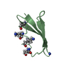
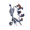
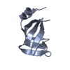
- PDB-5d0i: Structure of RING finger protein 165 -
+
Open data
ID or keywords:
Loading...
-
Basic information
Entry
Database: PDB / ID: 5d0i
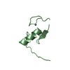
Title
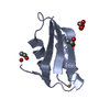
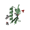
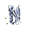
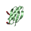
Structure of RING finger protein 165
Components
RING finger protein 165
Keywords
METAL BINDING PROTEIN / RING finger protein
Function / homology
Function and homology information
muscle structure development / forelimb morphogenesis / positive regulation of BMP signaling pathway / motor neuron axon guidance / innervation / protein catabolic process / RING-type E3 ubiquitin transferase / protein polyubiquitination / ubiquitin protein ligase activity / protein-containing complex ...muscle structure development / forelimb morphogenesis / positive regulation of BMP signaling pathway / motor neuron axon guidance / innervation / protein catabolic process / RING-type E3 ubiquitin transferase / protein polyubiquitination / ubiquitin protein ligase activity / protein-containing complex / zinc ion binding / nucleus Similarity search - Function
E3 ubiquitin-protein ligase MBR1/2-like / Zinc finger, RING-CH-type / The RING-variant domain is a C4HC3 zinc-finger like motif found in a number of cellular and viral proteins. Some of these proteins have been shown both in vivo and in vitro to have ubiquitin E3 ligase activity. / Ring finger domain / Zinc/RING finger domain, C3HC4 (zinc finger) / Herpes Virus-1 / Ring finger / Zinc finger RING-type profile. / Zinc finger, RING-type / Zinc finger, RING/FYVE/PHD-type ...E3 ubiquitin-protein ligase MBR1/2-like / Zinc finger, RING-CH-type / The RING-variant domain is a C4HC3 zinc-finger like motif found in a number of cellular and viral proteins. Some of these proteins have been shown both in vivo and in vitro to have ubiquitin E3 ligase activity. / Ring finger domain / Zinc/RING finger domain, C3HC4 (zinc finger) / Herpes Virus-1 / Ring finger / Zinc finger RING-type profile. / Zinc finger, RING-type / Zinc finger, RING/FYVE/PHD-type / 2-Layer Sandwich / Alpha Beta Similarity search - Domain/homology
In the structure databanks used in Yorodumi, some data are registered as the other names, "COVID-19 virus" and "2019-nCoV". Here are the details of the virus and the list of structure data.
Jan 31, 2019. EMDB accession codes are about to change! (news from PDBe EMDB page)
EMDB accession codes are about to change! (news from PDBe EMDB page)
The allocation of 4 digits for EMDB accession codes will soon come to an end. Whilst these codes will remain in use, new EMDB accession codes will include an additional digit and will expand incrementally as the available range of codes is exhausted. The current 4-digit format prefixed with “EMD-” (i.e. EMD-XXXX) will advance to a 5-digit format (i.e. EMD-XXXXX), and so on. It is currently estimated that the 4-digit codes will be depleted around Spring 2019, at which point the 5-digit format will come into force.
The EM Navigator/Yorodumi systems omit the EMD- prefix.
Related info.:Q: What is EMD? / ID/Accession-code notation in Yorodumi/EM Navigator
Yorodumi is a browser for structure data from EMDB, PDB, SASBDB, etc.
This page is also the successor to EM Navigator detail page, and also detail information page/front-end page for Omokage search.
The word "yorodu" (or yorozu) is an old Japanese word meaning "ten thousand". "mi" (miru) is to see.
Related info.:EMDB / PDB / SASBDB / Comparison of 3 databanks / Yorodumi Search / Aug 31, 2016. New EM Navigator & Yorodumi / Yorodumi Papers / Jmol/JSmol / Function and homology information / Changes in new EM Navigator and Yorodumi
 Movie
Movie Controller


 Open data
 Basic information
 Components
 Keywords
 Function and homology information
 Homo sapiens (human)
 X-RAY DIFFRACTION /
  Authors
 Citation
 Structure visualization
 Downloads & links
 Other downloads

 PDBj
PDBj

 Assembly
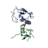
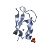


 Mass: 65.409 Da / Num. of mol.: 4 / Source method: obtained synthetically / Formula: Zn
 Mass: 96.063 Da / Num. of mol.: 1 / Source method: obtained synthetically / Formula: SO4
 Mass: 18.015 Da / Num. of mol.: 65 / Source method: isolated from a natural source / Formula: H2O
 Sample preparation
 / Beamline: MX1 / Wavelength: 0.9537 Å
 Processing