 Movie
Movie Controller

[English] 日本語
 Yorodumi
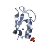
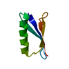
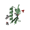
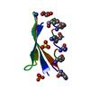
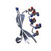
- PDB-2n7j: Sidechain chi1 distribution in B3 domain of protein G from extens... -
+ Open data

- Basic information
| Entry | Database: PDB / ID: 2n7j | ||||||
|---|---|---|---|---|---|---|---|
| Title | Sidechain chi1 distribution in B3 domain of protein G from extensive sets of residual dipolar couplings | ||||||
 Components Components | Immunoglobulin G-binding protein G | ||||||
 Keywords Keywords | SIGNALING PROTEIN | ||||||
| Function / homology |  Function and homology information Function and homology information | ||||||
| Biological species |  Streptococcus sp. 'group G' (bacteria) Streptococcus sp. 'group G' (bacteria) | ||||||
| Method | SOLUTION NMR / simulated annealing | ||||||
 Authors Authors | Grishaev, A. / Li, F. / Ying, J. / Bax, A. | ||||||
 Citation Citation | Journal: J.Am.Chem.Soc. / Year: 2015 Title: Side Chain Conformational Distributions of a Small Protein Derived from Model-Free Analysis of a Large Set of Residual Dipolar Couplings. Authors: Li, F. / Grishaev, A. / Ying, J. / Bax, A. | ||||||
| History |
|
- Structure visualization
| Structure viewer | Molecule: MolmilJmol/JSmol |
|---|
- Downloads & links
-Download
| PDBx/mmCIF format | 2n7j.cif.gz | 337.3 KB | Display | PDBx/mmCIF format |
|---|---|---|---|---|
| PDB format | pdb2n7j.ent.gz | 281.2 KB | Display | PDB format |
| PDBx/mmJSON format | 2n7j.json.gz | Tree view | PDBx/mmJSON format | |
| Others |  Other downloads Other downloads |
-Validation report
| Arichive directory | https://data.pdbj.org/pub/pdb/validation_reports/n7/2n7jftp://data.pdbj.org/pub/pdb/validation_reports/n7/2n7j | HTTPS FTP |
|---|
-Related structure data
| Related structure data | |
|---|---|
| Similar structure data | |
| Other databases |


-Links
 PDBj
PDBj

- Assembly
| Deposited unit | 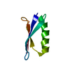
| |||||||||
|---|---|---|---|---|---|---|---|---|---|---|
| 1 |
| |||||||||
| NMR ensembles |
|
-Components
| #1: Antibody | Mass: 6214.848 Da / Num. of mol.: 1 / Fragment: residues 299-352 Source method: isolated from a genetically manipulated source Source: (gene. exp.) Streptococcus sp. 'group G' (bacteria) / Gene: spg / Production host: |
|---|
-Experimental details
-Experiment
| Experiment | Method: SOLUTION NMR Details: The backbone structure of the B3 domain of protein G is obtained as a single-conformation fit of an extensive set of backbone and Ca-Cb RDCs measured under multiple alignment orientations. ...Details: The backbone structure of the B3 domain of protein G is obtained as a single-conformation fit of an extensive set of backbone and Ca-Cb RDCs measured under multiple alignment orientations. Sidechain chi1 angle distributions are obtained from an ensemble fit to the experimental sidechain RDCs measured under six alignment orientations. Fitted data included separate CB-HB2 and CB-HB3 RDCs when available, as well as their sums (measured separately) as well as Cb-Cg RDCs where available. | ||||||||||||||||||||||||||||||||||||||||||||||||||||||||||||||||||||||||||||||||||||||||||||||||||||||||||||||||||||
|---|---|---|---|---|---|---|---|---|---|---|---|---|---|---|---|---|---|---|---|---|---|---|---|---|---|---|---|---|---|---|---|---|---|---|---|---|---|---|---|---|---|---|---|---|---|---|---|---|---|---|---|---|---|---|---|---|---|---|---|---|---|---|---|---|---|---|---|---|---|---|---|---|---|---|---|---|---|---|---|---|---|---|---|---|---|---|---|---|---|---|---|---|---|---|---|---|---|---|---|---|---|---|---|---|---|---|---|---|---|---|---|---|---|---|---|---|---|
| NMR experiment |
| ||||||||||||||||||||||||||||||||||||||||||||||||||||||||||||||||||||||||||||||||||||||||||||||||||||||||||||||||||||
| NMR details | Text: MODEL 1 SHOWS THE SIDECHAIN CONFORMERS IN THEIR MOST POPULATED CONFORMATIONS. MODELS 2-20 SHOW THE STATISTICAL DISTRIBUTION OF SIDECHAIN CONFORMERS CONSISTENT WITH THE SIDECHAIN RDC DATA. NOTE ...Text: MODEL 1 SHOWS THE SIDECHAIN CONFORMERS IN THEIR MOST POPULATED CONFORMATIONS. MODELS 2-20 SHOW THE STATISTICAL DISTRIBUTION OF SIDECHAIN CONFORMERS CONSISTENT WITH THE SIDECHAIN RDC DATA. NOTE THAT EXCEPT FOR RESIDUES POPULATING A SINGLE NARROWLY DISTRIBUTED RANGE OF CHI1 VALUES, SIDECHAIN RDCS ARE INCONSISTENT WITH ANY OF THE INDIVIDUAL MODELS. |
 HSQC
HSQC- Sample preparation
| Details |
| ||||||||||||||||||||||||||||||||||||||||||||||||||||||||||||||||||||
|---|---|---|---|---|---|---|---|---|---|---|---|---|---|---|---|---|---|---|---|---|---|---|---|---|---|---|---|---|---|---|---|---|---|---|---|---|---|---|---|---|---|---|---|---|---|---|---|---|---|---|---|---|---|---|---|---|---|---|---|---|---|---|---|---|---|---|---|---|---|
| Sample |
| ||||||||||||||||||||||||||||||||||||||||||||||||||||||||||||||||||||
| Sample conditions | Ionic strength: 0.05 / pH: 6.5 / Pressure: ambient / Temperature: 293 K |
-NMR measurement
| NMR spectrometer |
|
|---|
- Processing
| NMR software |
| ||||||||||||
|---|---|---|---|---|---|---|---|---|---|---|---|---|---|
| Refinement | Method: simulated annealing / Software ordinal: 1 Details: STRUCTURES WERE OBTAINED BY A SINGLE-CONFORMER SIMULATED ANNEALING REFINEMENT WITH VARIABLE ALIGNMENT TENSORS AGAINST AN EXTENSIVE SET OF BACKBONE AND CA-CB RDCS IN MULTIPLE ALIGNMENT MEDIA, ...Details: STRUCTURES WERE OBTAINED BY A SINGLE-CONFORMER SIMULATED ANNEALING REFINEMENT WITH VARIABLE ALIGNMENT TENSORS AGAINST AN EXTENSIVE SET OF BACKBONE AND CA-CB RDCS IN MULTIPLE ALIGNMENT MEDIA, J-COUPLINGS INCLUDING HN-HA, C'-C', AND THROUGH HYDROGEN BOND N-C', AND A BACKBONE-BACKBONE HYDROGEN BONDING DATABASE-DERIVED POTENTIAL OF MEAN FORCE. BACKBONE GEOMETRIES RESULTING FROM THIS STEP WERE KEPT FIXED UP TO CB ATOMS AND CHI1 TORSION ANGLES WERE RANDOMLY SELECTED TO SAMPLE THE DISTRIBUTIONS DERIVED FROM THE ANALYSIS OF AN EXTENSIVE DATASET OF CB-CG AND CB-HB RDCS IN 6 ALIGNMENT MEDIA. CLASHING WAS MINIMIZED BY RANDOMLY SAMPLING CHI2, CHI3 AND CHI4 TORSION ANGLES SELECTED FROM THE KINEMAGE DATABASE. MODELS 2-20 SHOWING MINIMAL CLASHING WERE THEN SELECTED FOR DEPOSITION. | ||||||||||||
| NMR constraints | Hydrogen bond constraints total count: 34 | ||||||||||||
| NMR representative | Selection criteria: sidechain conformers in their most populated conformations | ||||||||||||
| NMR ensemble | Conformer selection criteria: structures with favorable non-bond energy Conformers calculated total number: 100 / Conformers submitted total number: 20 |
