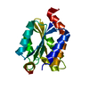
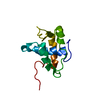
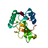
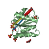
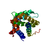
JC Virus large T-antigen origin binding domain F258L mutant
Components
Large T antigen
Keywords
viral protein / dna binding protein / DNA binding domain / origin binding domain
Function / homology
Function and homology information
symbiont-mediated suppression of host JAK-STAT cascade via inhibition of JAK1 activity / symbiont-mediated perturbation of host cell cycle G1/S transition checkpoint / 3'-5' DNA helicase activity / DNA 3'-5' helicase / DNA replication origin binding / DNA replication / symbiont-mediated suppression of host innate immune response / symbiont-mediated suppression of host type I interferon-mediated signaling pathway / host cell nucleus / ATP hydrolysis activity ...symbiont-mediated suppression of host JAK-STAT cascade via inhibition of JAK1 activity / symbiont-mediated perturbation of host cell cycle G1/S transition checkpoint / 3'-5' DNA helicase activity / DNA 3'-5' helicase / DNA replication origin binding / DNA replication / symbiont-mediated suppression of host innate immune response / symbiont-mediated suppression of host type I interferon-mediated signaling pathway / host cell nucleus / ATP hydrolysis activity / zinc ion binding / ATP binding Similarity search - Function
Replication Protein E1; Chain: A, - #20 / Replication Protein E1; Chain: A, / Large T antigen, polyomaviridae / Origin of replication binding protein / Large T-antigen (T-ag) origin-binding domain (OBD) profile. / T antigen, Ori-binding / Large T antigen, polyomavirus, C-terminal / Zinc finger, large T-antigen D1-type / Polyomavirus large T antigen C-terminus / Zinc finger large T-antigen (T-ag) D1-type profile. ...Replication Protein E1; Chain: A, - #20 / Replication Protein E1; Chain: A, / Large T antigen, polyomaviridae / Origin of replication binding protein / Large T-antigen (T-ag) origin-binding domain (OBD) profile. / T antigen, Ori-binding / Large T antigen, polyomavirus, C-terminal / Zinc finger, large T-antigen D1-type / Polyomavirus large T antigen C-terminus / Zinc finger large T-antigen (T-ag) D1-type profile. / Zinc finger, large T-antigen D1 domain superfamily / Helicase, superfamily 3, DNA virus / Superfamily 3 helicase of DNA viruses domain profile. / DnaJ molecular chaperone homology domain / dnaJ domain profile. / Chaperone J-domain superfamily / DnaJ domain / P-loop containing nucleoside triphosphate hydrolase / 3-Layer(aba) Sandwich / Alpha Beta Similarity search - Domain/homology
Resolution: 2.7→50 Å / Cor.coef. Fo:Fc: 0.949 / Cor.coef. Fo:Fc free: 0.939 / SU B: 23.55 / SU ML: 0.218 / Cross valid method: THROUGHOUT / ESU R: 0.63 / ESU R Free: 0.288 / Stereochemistry target values: MAXIMUM LIKELIHOOD / Details: HYDROGENS HAVE BEEN ADDED IN THE RIDING POSITIONS
Rfactor
Num. reflection
% reflection
Selection details
Rfree
0.22747
263
5.1 %
RANDOM
Rwork
0.1952
-
-
-
obs
0.19682
4927
99.98 %
-
Solvent computation
Ion probe radii: 0.8 Å / Shrinkage radii: 0.8 Å / VDW probe radii: 1 Å / Solvent model: MASK
 Movie
Movie Controller


 Open data
 Basic information
 Components
 Keywords
 Function and homology information
 JC polyomavirus
 X-RAY DIFFRACTION /
  Authors
 Citation
 Structure visualization
 Downloads & links
 Other downloads
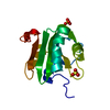


 PDBj
PDBj

 Assembly


 Mass: 62.068 Da / Num. of mol.: 1 / Source method: obtained synthetically / Formula: C2H6O2
 Mass: 18.015 Da / Num. of mol.: 7 / Source method: isolated from a natural source / Formula: H2O
 Sample preparation
 / Beamline: X29A / Wavelength: 1.075 Å
 Processing