 Movie
Movie Controller

[English] 日本語
 Yorodumi
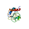
- PDB-4lif: Crystal structure of the JCV large T-antigen origin binding domain -
+ Open data

- Basic information
| Entry | Database: PDB / ID: 4lif | ||||||
|---|---|---|---|---|---|---|---|
| Title | Crystal structure of the JCV large T-antigen origin binding domain | ||||||
 Components Components | Large T antigen | ||||||
 Keywords Keywords | Hydrolase / dna binding protein / origin binding domain / replication / polyomavirus | ||||||
| Function / homology |  Function and homology information Function and homology informationsymbiont-mediated suppression of host JAK-STAT cascade via inhibition of JAK1 activity / symbiont-mediated perturbation of host cell cycle G1/S transition checkpoint / 3'-5' DNA helicase activity / DNA 3'-5' helicase / DNA replication origin binding / DNA replication / symbiont-mediated suppression of host innate immune response / symbiont-mediated suppression of host type I interferon-mediated signaling pathway / host cell nucleus / ATP hydrolysis activity ...symbiont-mediated suppression of host JAK-STAT cascade via inhibition of JAK1 activity / symbiont-mediated perturbation of host cell cycle G1/S transition checkpoint / 3'-5' DNA helicase activity / DNA 3'-5' helicase / DNA replication origin binding / DNA replication / symbiont-mediated suppression of host innate immune response / symbiont-mediated suppression of host type I interferon-mediated signaling pathway / host cell nucleus / ATP hydrolysis activity / zinc ion binding / ATP binding Similarity search - Function | ||||||
| Biological species |   JC polyomavirus JC polyomavirus | ||||||
| Method |  X-RAY DIFFRACTION / SYNCHROTRON / MOLECULAR REPLACEMENT / Resolution: 2.6 Å X-RAY DIFFRACTION / SYNCHROTRON / MOLECULAR REPLACEMENT / Resolution: 2.6 Å | ||||||
 Authors Authors | Meinke, G. / Bohm, A. / Bullock, P. | ||||||
 Citation Citation | Journal: Plos Pathog. / Year: 2014 Title: Insights into the Initiation of JC Virus DNA Replication Derived from the Crystal Structure of the T-Antigen Origin Binding Domain. Authors: Meinke, G. / Phelan, P.J. / Kalekar, R. / Shin, J. / Archambault, J. / Bohm, A. / Bullock, P.A. | ||||||
| History |
|
- Structure visualization
| Structure viewer | Molecule: MolmilJmol/JSmol |
|---|
- Downloads & links
-Download
| PDBx/mmCIF format | 4lif.cif.gz | 66.4 KB | Display | PDBx/mmCIF format |
|---|---|---|---|---|
| PDB format | pdb4lif.ent.gz | 48.9 KB | Display | PDB format |
| PDBx/mmJSON format | 4lif.json.gz | Tree view | PDBx/mmJSON format | |
| Others |  Other downloads Other downloads |
-Validation report
| Arichive directory | https://data.pdbj.org/pub/pdb/validation_reports/li/4lifftp://data.pdbj.org/pub/pdb/validation_reports/li/4lif | HTTPS FTP |
|---|
-Related structure data
| Related structure data | 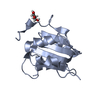 4lmdC  4nbpC 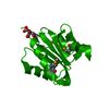 2fufS C: citing same article ( S: Starting model for refinement |
|---|---|
| Similar structure data |


-Links
 PDBj
PDBj

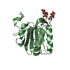
- Assembly
| Deposited unit | 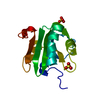
| ||||||||
|---|---|---|---|---|---|---|---|---|---|
| 1 |
| ||||||||
| Unit cell |
|
-Components
| #1: Protein | Mass: 15170.390 Da / Num. of mol.: 1 / Fragment: OBD (unp residues 132-261) Source method: isolated from a genetically manipulated source Source: (gene. exp.) JC polyomavirus / Gene: Large T antigen / Plasmid: pGEX-4T / Production host:  References: UniProt: P03072, Hydrolases; Acting on acid anhydrides; Acting on acid anhydrides to facilitate cellular and subcellular movement | ||
|---|---|---|---|
| #2: Chemical |   Mass: 96.063 Da / Num. of mol.: 2 / Source method: obtained synthetically / Formula: SO4 Mass: 96.063 Da / Num. of mol.: 2 / Source method: obtained synthetically / Formula: SO4#3: Water | ChemComp-HOH / |  Mass: 18.015 Da / Num. of mol.: 5 / Source method: isolated from a natural source / Formula: H2O Mass: 18.015 Da / Num. of mol.: 5 / Source method: isolated from a natural source / Formula: H2O |
-Experimental details
-Experiment
| Experiment | Method: X-RAY DIFFRACTION / Number of used crystals: 1 |
|---|
- Sample preparation
| Crystal | Density Matthews: 3.09 Å3/Da / Density % sol: 60.25 % |
|---|---|
| Crystal grow | Temperature: 273 K / Method: vapor diffusion, sitting drop / pH: 8.5 Details: 0.1 M Tris, 0.2 M LiSO4, 30 % Peg 4000, VAPOR DIFFUSION, SITTING DROP, temperature 273K, pH 8.5 |
-Data collection
| Diffraction | Mean temperature: 100 K |
|---|---|
| Diffraction source | Source: SYNCHROTRON / Site: NSLS  / Beamline: X29A / Wavelength: 1.075 Å / Beamline: X29A / Wavelength: 1.075 Å |
| Detector | Type: ADSC QUANTUM 315r / Detector: CCD / Date: Mar 14, 2013 |
| Radiation | Monochromator: Si(111) / Protocol: SINGLE WAVELENGTH / Monochromatic (M) / Laue (L): M / Scattering type: x-ray |
| Radiation wavelength | Wavelength: 1.075 Å / Relative weight: 1 |
| Reflection | Resolution: 2.6→50 Å / Num. all: 5909 / Num. obs: 5906 / % possible obs: 99.8 % / Observed criterion σ(F): 0 / Observed criterion σ(I): 0 / Redundancy: 14.7 % / Biso Wilson estimate: 72.4 Å2 / Rmerge(I) obs: 0.068 / Net I/σ(I): 37.6 |
- Processing
| Software |
| |||||||||||||||||||||||||||||||||||||||||||||||||||||||||||||||||||||||||||
|---|---|---|---|---|---|---|---|---|---|---|---|---|---|---|---|---|---|---|---|---|---|---|---|---|---|---|---|---|---|---|---|---|---|---|---|---|---|---|---|---|---|---|---|---|---|---|---|---|---|---|---|---|---|---|---|---|---|---|---|---|---|---|---|---|---|---|---|---|---|---|---|---|---|---|---|---|
| Refinement | Method to determine structure: MOLECULAR REPLACEMENT Starting model: PDB ENTRY 2FUF Resolution: 2.6→36.696 Å / SU ML: 0.22 / Cross valid method: THROUGHOUT / σ(F): 0.39 / Phase error: 25.57 / Stereochemistry target values: ML
| |||||||||||||||||||||||||||||||||||||||||||||||||||||||||||||||||||||||||||
| Solvent computation | Shrinkage radii: 0.9 Å / VDW probe radii: 1.11 Å / Solvent model: FLAT BULK SOLVENT MODEL | |||||||||||||||||||||||||||||||||||||||||||||||||||||||||||||||||||||||||||
| Refinement step | Cycle: LAST / Resolution: 2.6→36.696 Å
| |||||||||||||||||||||||||||||||||||||||||||||||||||||||||||||||||||||||||||
| Refine LS restraints |
| |||||||||||||||||||||||||||||||||||||||||||||||||||||||||||||||||||||||||||
| LS refinement shell |
| |||||||||||||||||||||||||||||||||||||||||||||||||||||||||||||||||||||||||||
| Refinement TLS params. | Method: refined / Refine-ID: X-RAY DIFFRACTION
| |||||||||||||||||||||||||||||||||||||||||||||||||||||||||||||||||||||||||||
| Refinement TLS group |
|
