 Movie
Movie Controller

[English] 日本語
 Yorodumi
- PDB-5cvs: GlgE isoform 1 from Streptomyces coelicolor E423A mutant soaked i... -
+ Open data

- Basic information
| Entry | Database: PDB / ID: 5cvs | ||||||||||||
|---|---|---|---|---|---|---|---|---|---|---|---|---|---|
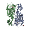
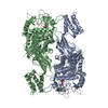
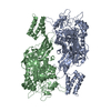
| Title | GlgE isoform 1 from Streptomyces coelicolor E423A mutant soaked in maltoheptaose | ||||||||||||
 Components Components | Alpha-1,4-glucan:maltose-1-phosphate maltosyltransferase 1 | ||||||||||||
 Keywords Keywords | TRANSFERASE / HYDROLASE / ALPHA-GLUCAN BIOSYNTHESIS / GLYCOSIDE HYDROLASE FAMILY 13_3 | ||||||||||||
| Function / homology |  Function and homology information Function and homology informationstarch synthase (maltosyl-transferring) / alpha-glucan biosynthetic process / hexosyltransferase activity / alpha-amylase activity / oligosaccharide catabolic process Similarity search - Function | ||||||||||||
| Biological species |  Streptomyces coelicolor (bacteria) Streptomyces coelicolor (bacteria) | ||||||||||||
| Method |  X-RAY DIFFRACTION / SYNCHROTRON / MOLECULAR REPLACEMENT / Resolution: 2.3 Å X-RAY DIFFRACTION / SYNCHROTRON / MOLECULAR REPLACEMENT / Resolution: 2.3 Å | ||||||||||||
 Authors Authors | Rashid, A.M. / Syson, K. / Koliwer-Brandl, H. / van de Weerd, R. / Stevenson, C.E.M. / Batey, S.F.D. / Miah, F. / Alber, M. / Ioerger, T.R. / Chandra, G. ...Rashid, A.M. / Syson, K. / Koliwer-Brandl, H. / van de Weerd, R. / Stevenson, C.E.M. / Batey, S.F.D. / Miah, F. / Alber, M. / Ioerger, T.R. / Chandra, G. / Appelmelk, B.J. / Nartowski, K.P. / Khimyak, Y.Z. / Lawson, D.M. / Jacobs, W.R. / Geurtsen, J. / Kalscheuer, R. / Bornemann, S. | ||||||||||||
| Funding support |  United Kingdom, United Kingdom,  Netherlands, 3items Netherlands, 3items
| ||||||||||||
 Citation Citation | Journal: J.Biol.Chem. / Year: 2016 Title: Ligand-bound structures and site-directed mutagenesis identify the acceptor and secondary binding sites of Streptomyces coelicolor maltosyltransferase GlgE. Authors: Syson, K. / Stevenson, C.E. / Miah, F. / Barclay, J.E. / Tang, M. / Gorelik, A. / Rashid, A.M. / Lawson, D.M. / Bornemann, S. | ||||||||||||
| History |
|
- Structure visualization
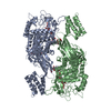
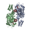
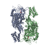
| Structure viewer | Molecule: MolmilJmol/JSmol |
|---|
- Downloads & links
-Download
| PDBx/mmCIF format | 5cvs.cif.gz | 533.7 KB | Display | PDBx/mmCIF format |
|---|---|---|---|---|
| PDB format | pdb5cvs.ent.gz | 441.7 KB | Display | PDB format |
| PDBx/mmJSON format | 5cvs.json.gz | Tree view | PDBx/mmJSON format | |
| Others |  Other downloads Other downloads |
-Validation report
| Arichive directory | https://data.pdbj.org/pub/pdb/validation_reports/cv/5cvsftp://data.pdbj.org/pub/pdb/validation_reports/cv/5cvs | HTTPS FTP |
|---|
-Related structure data
| Related structure data |  5lgvC 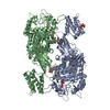 5lgwC  4cn6S S: Starting model for refinement C: citing same article ( |
|---|---|
| Similar structure data |


-Links
 PDBj
PDBj


- Assembly
| Deposited unit | 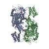
| ||||||||||||||||||
|---|---|---|---|---|---|---|---|---|---|---|---|---|---|---|---|---|---|---|---|
| 1 |
| ||||||||||||||||||
| Unit cell |
| ||||||||||||||||||
| Noncrystallographic symmetry (NCS) | NCS domain:
NCS domain segments: Component-ID: _ / Ens-ID: 1 / Beg auth comp-ID: PRO / Beg label comp-ID: PRO / End auth comp-ID: ARG / End label comp-ID: ARG / Refine code: _ / Auth seq-ID: 15 - 663 / Label seq-ID: 15 - 663
|
-Components
| #1: Protein | Mass: 75335.438 Da / Num. of mol.: 2 / Mutation: E423A Source method: isolated from a genetically manipulated source Source: (gene. exp.) Streptomyces coelicolor (bacteria) / Gene: glgE1, pep1, pep1A, pep1I, SCO5443, SC6A11.19c / Plasmid: PET15B / Details (production host): PET15B-GLGE1-M145 / Production host: References: UniProt: Q9L1K2, starch synthase (maltosyl-transferring) #2: Polysaccharide | Source method: isolated from a genetically manipulated source #3: Polysaccharide | 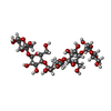  Source method: isolated from a genetically manipulated source Details: oligosaccharide / References: alpha-maltopentaose #4: Water | ChemComp-HOH / |  Mass: 18.015 Da / Num. of mol.: 484 / Source method: isolated from a natural source / Formula: H2O Mass: 18.015 Da / Num. of mol.: 484 / Source method: isolated from a natural source / Formula: H2O |
|---|
-Experimental details
-Experiment
| Experiment | Method: X-RAY DIFFRACTION / Number of used crystals: 1 |
|---|
- Sample preparation
| Crystal | Density Matthews: 3.39 Å3/Da / Density % sol: 63.74 % |
|---|---|
| Crystal grow | Temperature: 293 K / Method: vapor diffusion, hanging drop / pH: 7 / Details: NULL |
-Data collection
| Diffraction | Mean temperature: 100 K | |||||||||||||||||||||||||||
|---|---|---|---|---|---|---|---|---|---|---|---|---|---|---|---|---|---|---|---|---|---|---|---|---|---|---|---|---|
| Diffraction source | Source: SYNCHROTRON / Site: Diamond / Beamline: I04-1 / Wavelength: 0.9173 Å | |||||||||||||||||||||||||||
| Detector | Type: DECTRIS PILATUS 2M / Detector: PIXEL / Date: Aug 1, 2014 | |||||||||||||||||||||||||||
| Radiation | Protocol: SINGLE WAVELENGTH / Monochromatic (M) / Laue (L): M / Scattering type: x-ray | |||||||||||||||||||||||||||
| Radiation wavelength | Wavelength: 0.9173 Å / Relative weight: 1 | |||||||||||||||||||||||||||
| Reflection | Resolution: 2.3→62.83 Å / Num. obs: 92902 / % possible obs: 99.9 % / Redundancy: 18.4 % / Biso Wilson estimate: 47 Å2 / CC1/2: 0.999 / Rmerge(I) obs: 0.069 / Rpim(I) all: 0.016 / Net I/σ(I): 27.6 / Num. measured all: 1708887 | |||||||||||||||||||||||||||
| Reflection shell | Diffraction-ID: 1 / Rejects: _
|
- Processing
| Software |
| |||||||||||||||||||||||||||||||||||||||||||||||||||||||||||||||||||||||||||||||||||||||||||||||||||||||||||||||||||||||||||||||||||||||||||||||||||||||||||||||||||||||||||||||||||||||||||||||||||||||||||||||||||||||||||||||||
|---|---|---|---|---|---|---|---|---|---|---|---|---|---|---|---|---|---|---|---|---|---|---|---|---|---|---|---|---|---|---|---|---|---|---|---|---|---|---|---|---|---|---|---|---|---|---|---|---|---|---|---|---|---|---|---|---|---|---|---|---|---|---|---|---|---|---|---|---|---|---|---|---|---|---|---|---|---|---|---|---|---|---|---|---|---|---|---|---|---|---|---|---|---|---|---|---|---|---|---|---|---|---|---|---|---|---|---|---|---|---|---|---|---|---|---|---|---|---|---|---|---|---|---|---|---|---|---|---|---|---|---|---|---|---|---|---|---|---|---|---|---|---|---|---|---|---|---|---|---|---|---|---|---|---|---|---|---|---|---|---|---|---|---|---|---|---|---|---|---|---|---|---|---|---|---|---|---|---|---|---|---|---|---|---|---|---|---|---|---|---|---|---|---|---|---|---|---|---|---|---|---|---|---|---|---|---|---|---|---|---|---|---|---|---|---|---|---|---|---|---|---|---|---|---|---|---|
| Refinement | Method to determine structure: MOLECULAR REPLACEMENT Starting model: 4CN6 Resolution: 2.3→62.83 Å / Cor.coef. Fo:Fc: 0.957 / Cor.coef. Fo:Fc free: 0.952 / WRfactor Rfree: 0.2012 / WRfactor Rwork: 0.1725 / FOM work R set: 0.8703 / SU B: 10.055 / SU ML: 0.127 / SU R Cruickshank DPI: 0.211 / SU Rfree: 0.1728 / Cross valid method: THROUGHOUT / σ(F): 0 / ESU R: 0.211 / ESU R Free: 0.173 / Stereochemistry target values: MAXIMUM LIKELIHOOD Details: HYDROGENS HAVE BEEN ADDED IN THE RIDING POSITIONS U VALUES : WITH TLS ADDED
| |||||||||||||||||||||||||||||||||||||||||||||||||||||||||||||||||||||||||||||||||||||||||||||||||||||||||||||||||||||||||||||||||||||||||||||||||||||||||||||||||||||||||||||||||||||||||||||||||||||||||||||||||||||||||||||||||
| Solvent computation | Ion probe radii: 0.8 Å / Shrinkage radii: 0.8 Å / VDW probe radii: 1.2 Å / Solvent model: MASK | |||||||||||||||||||||||||||||||||||||||||||||||||||||||||||||||||||||||||||||||||||||||||||||||||||||||||||||||||||||||||||||||||||||||||||||||||||||||||||||||||||||||||||||||||||||||||||||||||||||||||||||||||||||||||||||||||
| Displacement parameters | Biso max: 139.36 Å2 / Biso mean: 59.9 Å2 / Biso min: 32.64 Å2
| |||||||||||||||||||||||||||||||||||||||||||||||||||||||||||||||||||||||||||||||||||||||||||||||||||||||||||||||||||||||||||||||||||||||||||||||||||||||||||||||||||||||||||||||||||||||||||||||||||||||||||||||||||||||||||||||||
| Refinement step | Cycle: final / Resolution: 2.3→62.83 Å
| |||||||||||||||||||||||||||||||||||||||||||||||||||||||||||||||||||||||||||||||||||||||||||||||||||||||||||||||||||||||||||||||||||||||||||||||||||||||||||||||||||||||||||||||||||||||||||||||||||||||||||||||||||||||||||||||||
| Refine LS restraints |
| |||||||||||||||||||||||||||||||||||||||||||||||||||||||||||||||||||||||||||||||||||||||||||||||||||||||||||||||||||||||||||||||||||||||||||||||||||||||||||||||||||||||||||||||||||||||||||||||||||||||||||||||||||||||||||||||||
| Refine LS restraints NCS | Ens-ID: 1 / Number: 75692 / Refine-ID: X-RAY DIFFRACTION / Type: interatomic distance / Rms dev position: 0.06 Å / Weight position: 0.05
| |||||||||||||||||||||||||||||||||||||||||||||||||||||||||||||||||||||||||||||||||||||||||||||||||||||||||||||||||||||||||||||||||||||||||||||||||||||||||||||||||||||||||||||||||||||||||||||||||||||||||||||||||||||||||||||||||
| LS refinement shell | Resolution: 2.3→2.36 Å / Total num. of bins used: 20
| |||||||||||||||||||||||||||||||||||||||||||||||||||||||||||||||||||||||||||||||||||||||||||||||||||||||||||||||||||||||||||||||||||||||||||||||||||||||||||||||||||||||||||||||||||||||||||||||||||||||||||||||||||||||||||||||||
| Refinement TLS params. | Method: refined / Refine-ID: X-RAY DIFFRACTION
| |||||||||||||||||||||||||||||||||||||||||||||||||||||||||||||||||||||||||||||||||||||||||||||||||||||||||||||||||||||||||||||||||||||||||||||||||||||||||||||||||||||||||||||||||||||||||||||||||||||||||||||||||||||||||||||||||
| Refinement TLS group |
|
