 Movie
Movie Controller


+ Open data

- Basic information
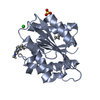
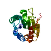
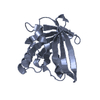
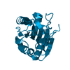
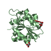
| Entry | Database: PDB / ID: 5ct6 | ||||||
|---|---|---|---|---|---|---|---|
| Title | Wild-type Bacillus subtilis lipase A with 20% [BMIM][Cl] | ||||||
 Components Components | Lipase EstA | ||||||
 Keywords Keywords | HYDROLASE | ||||||
| Function / homology |  Function and homology information Function and homology informationlipase activity / triacylglycerol lipase / triacylglycerol lipase activity / lipid catabolic process / extracellular region Similarity search - Function | ||||||
| Biological species |  | ||||||
| Method |  X-RAY DIFFRACTION / SYNCHROTRON / Resolution: 1.902 Å X-RAY DIFFRACTION / SYNCHROTRON / Resolution: 1.902 Å | ||||||
 Authors Authors | Nordwald, E.M. / Plaks, J.G. / Snell, J.R. / Sousa, M.C. / Kaar, J.L. | ||||||
 Citation Citation | Journal: Chembiochem / Year: 2015 Title: Crystallographic Investigation of Imidazolium Ionic Liquid Effects on Enzyme Structure. Authors: Nordwald, E.M. / Plaks, J.G. / Snell, J.R. / Sousa, M.C. / Kaar, J.L. | ||||||
| History |
|
- Structure visualization
| Structure viewer | Molecule: MolmilJmol/JSmol |
|---|
- Downloads & links
-Download
| PDBx/mmCIF format | 5ct6.cif.gz | 151.4 KB | Display | PDBx/mmCIF format |
|---|---|---|---|---|
| PDB format | pdb5ct6.ent.gz | 125.8 KB | Display | PDB format |
| PDBx/mmJSON format | 5ct6.json.gz | Tree view | PDBx/mmJSON format | |
| Others |  Other downloads Other downloads |
-Validation report
| Arichive directory | https://data.pdbj.org/pub/pdb/validation_reports/ct/5ct6ftp://data.pdbj.org/pub/pdb/validation_reports/ct/5ct6 | HTTPS FTP |
|---|
-Related structure data
| Related structure data | 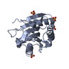 5criC  5ct4C 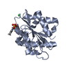 5ct5C 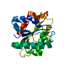 5ct8C  5ct9C 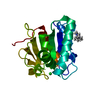 5ctaC 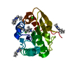 5curC C: citing same article ( |
|---|---|
| Similar structure data |


-Links
 PDBj
PDBj- Assembly
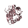
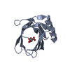
| Deposited unit | 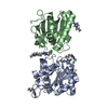
| ||||||||
|---|---|---|---|---|---|---|---|---|---|
| 1 | 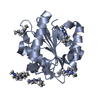
| ||||||||
| 2 | 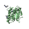
| ||||||||
| 3 |
| ||||||||
| Unit cell |
|
-Components
| #1: Protein | Mass: 19311.758 Da / Num. of mol.: 2 Source method: isolated from a genetically manipulated source Source: (gene. exp.) Strain: 168 / Gene: estA, lip, lipA, BSU02700 / Plasmid: pET21b / Production host: #2: Chemical |   Mass: 35.453 Da / Num. of mol.: 3 / Source method: obtained synthetically / Formula: Cl Mass: 35.453 Da / Num. of mol.: 3 / Source method: obtained synthetically / Formula: Cl#3: Chemical | ChemComp-BM0 / 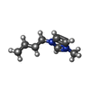  Mass: 139.218 Da / Num. of mol.: 8 / Source method: obtained synthetically / Formula: C8H15N2 Mass: 139.218 Da / Num. of mol.: 8 / Source method: obtained synthetically / Formula: C8H15N2#4: Water | ChemComp-HOH / |  Mass: 18.015 Da / Num. of mol.: 232 / Source method: isolated from a natural source / Formula: H2O Mass: 18.015 Da / Num. of mol.: 232 / Source method: isolated from a natural source / Formula: H2O |
|---|
-Experimental details
-Experiment
| Experiment | Method: X-RAY DIFFRACTION |
|---|
- Sample preparation
| Crystal | Density Matthews: 2.05 Å3/Da / Density % sol: 40.11 % |
|---|---|
| Crystal grow | Temperature: 295 K / Method: vapor diffusion, hanging drop Details: 35 % PEG 3350, 20 mM NaSO4, 0.1M ethanolamine, 10mM ZnCl2, pH 9.5 |
-Data collection
| Diffraction | Mean temperature: 100 K |
|---|---|
| Diffraction source | Source: SYNCHROTRON / Site: ALS  / Beamline: 8.2.2 / Wavelength: 0.9999 Å / Beamline: 8.2.2 / Wavelength: 0.9999 Å |
| Detector | Type: ADSC QUANTUM 315 / Detector: CCD / Date: Dec 13, 2014 |
| Radiation | Protocol: SINGLE WAVELENGTH / Monochromatic (M) / Laue (L): M / Scattering type: x-ray |
| Radiation wavelength | Wavelength: 0.9999 Å / Relative weight: 1 |
| Reflection | Resolution: 1.9→48 Å / Num. obs: 25866 / % possible obs: 100 % / Redundancy: 3.7 % / Rmerge(I) obs: 0.122 / Net I/σ(I): 14.5 |
| Reflection shell | Resolution: 1.9→1.93 Å / Redundancy: 3.4 % / Rmerge(I) obs: 0 / Mean I/σ(I) obs: 1 / % possible all: 100 |
- Processing
| Software |
| |||||||||||||||||||||||||||||||||||||||||||||||||||||||||||||||||||||||||||||||||||||||||||||||||||||||||
|---|---|---|---|---|---|---|---|---|---|---|---|---|---|---|---|---|---|---|---|---|---|---|---|---|---|---|---|---|---|---|---|---|---|---|---|---|---|---|---|---|---|---|---|---|---|---|---|---|---|---|---|---|---|---|---|---|---|---|---|---|---|---|---|---|---|---|---|---|---|---|---|---|---|---|---|---|---|---|---|---|---|---|---|---|---|---|---|---|---|---|---|---|---|---|---|---|---|---|---|---|---|---|---|---|---|---|
| Refinement | Resolution: 1.902→47.907 Å / SU ML: 0.27 / Cross valid method: FREE R-VALUE / σ(F): 1.34 / Phase error: 21.33 / Stereochemistry target values: ML
| |||||||||||||||||||||||||||||||||||||||||||||||||||||||||||||||||||||||||||||||||||||||||||||||||||||||||
| Solvent computation | Shrinkage radii: 0.9 Å / VDW probe radii: 1.11 Å / Solvent model: FLAT BULK SOLVENT MODEL / Bsol: 37.139 Å2 / ksol: 0.331 e/Å3 | |||||||||||||||||||||||||||||||||||||||||||||||||||||||||||||||||||||||||||||||||||||||||||||||||||||||||
| Displacement parameters |
| |||||||||||||||||||||||||||||||||||||||||||||||||||||||||||||||||||||||||||||||||||||||||||||||||||||||||
| Refinement step | Cycle: LAST / Resolution: 1.902→47.907 Å
| |||||||||||||||||||||||||||||||||||||||||||||||||||||||||||||||||||||||||||||||||||||||||||||||||||||||||
| Refine LS restraints |
| |||||||||||||||||||||||||||||||||||||||||||||||||||||||||||||||||||||||||||||||||||||||||||||||||||||||||
| LS refinement shell |
|
