 Movie
Movie Controller


+ Open data

- Basic information
| Entry | Database: PDB / ID: 4run | ||||||
|---|---|---|---|---|---|---|---|
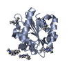
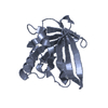
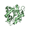
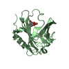
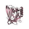
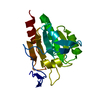
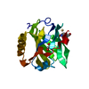
| Title | Crystal structure of human odorant binding protein OBPIIa | ||||||
 Components Components | Odorant-binding protein 2a | ||||||
 Keywords Keywords | TRANSPORT PROTEIN / human lipocalin / lipid binding protein / odorant binding protein / vanillin / lilial / fatty acids / aliphatic aldehydes / nasal mucus | ||||||
| Function / homology |  Function and homology information Function and homology informationodorant binding / sensory perception of chemical stimulus / small molecule binding / sensory perception of smell / glucose homeostasis / : Similarity search - Function | ||||||
| Biological species |  Homo sapiens (human) Homo sapiens (human) | ||||||
| Method |  X-RAY DIFFRACTION / SYNCHROTRON / MOLECULAR REPLACEMENT / Resolution: 2.6 Å X-RAY DIFFRACTION / SYNCHROTRON / MOLECULAR REPLACEMENT / Resolution: 2.6 Å | ||||||
 Authors Authors | Schiefner, A. / Skerra, A. | ||||||
 Citation Citation | Journal: Proteins / Year: 2015 Title: Crystal structure of the human odorant binding protein, OBPIIa. Authors: Schiefner, A. / Freier, R. / Eichinger, A. / Skerra, A. | ||||||
| History |
|
- Structure visualization
| Structure viewer | Molecule: MolmilJmol/JSmol |
|---|
- Downloads & links
-Download
| PDBx/mmCIF format | 4run.cif.gz | 136 KB | Display | PDBx/mmCIF format |
|---|---|---|---|---|
| PDB format | pdb4run.ent.gz | 107.5 KB | Display | PDB format |
| PDBx/mmJSON format | 4run.json.gz | Tree view | PDBx/mmJSON format | |
| Others |  Other downloads Other downloads |
-Validation report
| Arichive directory | https://data.pdbj.org/pub/pdb/validation_reports/ru/4runftp://data.pdbj.org/pub/pdb/validation_reports/ru/4run | HTTPS FTP |
|---|
-Related structure data
| Related structure data |  3eycS S: Starting model for refinement |
|---|---|
| Similar structure data |


-Links
 PDBj
PDBj


- Assembly
| Deposited unit | 
| ||||||||||||||||||
|---|---|---|---|---|---|---|---|---|---|---|---|---|---|---|---|---|---|---|---|
| 1 | 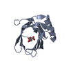
| ||||||||||||||||||
| 2 | 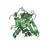
| ||||||||||||||||||
| 3 |
| ||||||||||||||||||
| Unit cell |
| ||||||||||||||||||
| Noncrystallographic symmetry (NCS) | NCS domain:
NCS domain segments: Component-ID: _ / Ens-ID: 1 / Beg auth comp-ID: GLU / Beg label comp-ID: GLU / End auth comp-ID: VAL / End label comp-ID: VAL / Refine code: _ / Auth seq-ID: 8 - 152 / Label seq-ID: 8 - 152
|
-Components
| #1: Protein | Mass: 18655.281 Da / Num. of mol.: 2 / Mutation: C99S, K112N Source method: isolated from a genetically manipulated source Source: (gene. exp.) Homo sapiens (human) / Gene: OBP2A / Plasmid: pASK75-his / Production host:  #2: Chemical | ChemComp-FLC / |   Mass: 189.100 Da / Num. of mol.: 1 / Source method: obtained synthetically / Formula: C6H5O7 Mass: 189.100 Da / Num. of mol.: 1 / Source method: obtained synthetically / Formula: C6H5O7#3: Water | ChemComp-HOH / |  Mass: 18.015 Da / Num. of mol.: 15 / Source method: isolated from a natural source / Formula: H2O Mass: 18.015 Da / Num. of mol.: 15 / Source method: isolated from a natural source / Formula: H2OHas protein modification | Y | |
|---|
-Experimental details
-Experiment
| Experiment | Method: X-RAY DIFFRACTION / Number of used crystals: 1 |
|---|
- Sample preparation
| Crystal | Density Matthews: 2.18 Å3/Da / Density % sol: 43.58 % |
|---|---|
| Crystal grow | Temperature: 293 K / Method: vapor diffusion, hanging drop / pH: 3.5 Details: Protein solution: 14 mg/ml protein, 0.2 M imidazole, 0.4 M NaCl, 10 mM Tris-HCl, 1.3 mM Menthol, 3.3 %(v/v) Methanol; Reservoir solution: 20 %(w/v) PEG 3350, 0.1 M ammonium citrate, pH 3.5, ...Details: Protein solution: 14 mg/ml protein, 0.2 M imidazole, 0.4 M NaCl, 10 mM Tris-HCl, 1.3 mM Menthol, 3.3 %(v/v) Methanol; Reservoir solution: 20 %(w/v) PEG 3350, 0.1 M ammonium citrate, pH 3.5, VAPOR DIFFUSION, HANGING DROP, temperature 293K |
-Data collection
| Diffraction | Mean temperature: 100 K | ||||||||||||||||||||||||||||||||||||||||||||||||||||||||||||||||||||||||||||||||||||
|---|---|---|---|---|---|---|---|---|---|---|---|---|---|---|---|---|---|---|---|---|---|---|---|---|---|---|---|---|---|---|---|---|---|---|---|---|---|---|---|---|---|---|---|---|---|---|---|---|---|---|---|---|---|---|---|---|---|---|---|---|---|---|---|---|---|---|---|---|---|---|---|---|---|---|---|---|---|---|---|---|---|---|---|---|---|
| Diffraction source | Source: SYNCHROTRON / Site: BESSY  / Beamline: 14.2 / Wavelength: 0.9184 Å / Beamline: 14.2 / Wavelength: 0.9184 Å | ||||||||||||||||||||||||||||||||||||||||||||||||||||||||||||||||||||||||||||||||||||
| Detector | Type: MARMOSAIC 225 mm CCD / Detector: CCD / Date: Dec 7, 2011 / Details: MIRRORS | ||||||||||||||||||||||||||||||||||||||||||||||||||||||||||||||||||||||||||||||||||||
| Radiation | Monochromator: SI-111 crystal / Protocol: SINGLE WAVELENGTH / Monochromatic (M) / Laue (L): M / Scattering type: x-ray | ||||||||||||||||||||||||||||||||||||||||||||||||||||||||||||||||||||||||||||||||||||
| Radiation wavelength | Wavelength: 0.9184 Å / Relative weight: 1 | ||||||||||||||||||||||||||||||||||||||||||||||||||||||||||||||||||||||||||||||||||||
| Reflection | Resolution: 2.6→35 Å / Num. all: 10926 / Num. obs: 10926 / % possible obs: 99.7 % / Observed criterion σ(F): -3 / Observed criterion σ(I): -3 / Redundancy: 8.8 % / Biso Wilson estimate: 63.91 Å2 / Rmerge(I) obs: 0.049 / Χ2: 0.985 / Net I/σ(I): 31.88 | ||||||||||||||||||||||||||||||||||||||||||||||||||||||||||||||||||||||||||||||||||||
| Reflection shell |
|
- Processing
| Software |
| |||||||||||||||||||||||||||||||||||||||||||||||||||||||||||||||||||||||||||
|---|---|---|---|---|---|---|---|---|---|---|---|---|---|---|---|---|---|---|---|---|---|---|---|---|---|---|---|---|---|---|---|---|---|---|---|---|---|---|---|---|---|---|---|---|---|---|---|---|---|---|---|---|---|---|---|---|---|---|---|---|---|---|---|---|---|---|---|---|---|---|---|---|---|---|---|---|
| Refinement | Method to determine structure: MOLECULAR REPLACEMENT Starting model: 3EYC Resolution: 2.6→33.7 Å / Cor.coef. Fo:Fc: 0.927 / Cor.coef. Fo:Fc free: 0.866 / WRfactor Rfree: 0.2651 / WRfactor Rwork: 0.2213 / FOM work R set: 0.7801 / SU B: 33.044 / SU ML: 0.328 / SU R Cruickshank DPI: 1.1842 / SU Rfree: 0.3639 / Cross valid method: THROUGHOUT / σ(F): 0 / ESU R: 1.184 / ESU R Free: 0.364 / Stereochemistry target values: MAXIMUM LIKELIHOOD Details: HYDROGENS HAVE BEEN ADDED IN THE RIDING POSITIONS U VALUES : WITH TLS ADDED
| |||||||||||||||||||||||||||||||||||||||||||||||||||||||||||||||||||||||||||
| Solvent computation | Ion probe radii: 0.8 Å / Shrinkage radii: 0.8 Å / VDW probe radii: 1.2 Å / Solvent model: MASK | |||||||||||||||||||||||||||||||||||||||||||||||||||||||||||||||||||||||||||
| Displacement parameters | Biso max: 174.07 Å2 / Biso mean: 75.6 Å2 / Biso min: 36.73 Å2
| |||||||||||||||||||||||||||||||||||||||||||||||||||||||||||||||||||||||||||
| Refinement step | Cycle: LAST / Resolution: 2.6→33.7 Å
| |||||||||||||||||||||||||||||||||||||||||||||||||||||||||||||||||||||||||||
| Refine LS restraints |
| |||||||||||||||||||||||||||||||||||||||||||||||||||||||||||||||||||||||||||
| Refine LS restraints NCS | Ens-ID: 1 / Number: 7513 / Refine-ID: X-RAY DIFFRACTION / Type: interatomic distance / Rms dev position: 0.16 Å / Weight position: 0.05
| |||||||||||||||||||||||||||||||||||||||||||||||||||||||||||||||||||||||||||
| LS refinement shell | Resolution: 2.6→2.667 Å / Total num. of bins used: 20
| |||||||||||||||||||||||||||||||||||||||||||||||||||||||||||||||||||||||||||
| Refinement TLS params. | Method: refined / Refine-ID: X-RAY DIFFRACTION
| |||||||||||||||||||||||||||||||||||||||||||||||||||||||||||||||||||||||||||
| Refinement TLS group |
|
