 Movie
Movie Controller

[English] 日本語
 Yorodumi
- PDB-5cir: Crystal structure of death receptor 4 (DR4; TNFFRSF10A) bound to ... -
+ Open data

- Basic information
| Entry | Database: PDB / ID: 5cir | ||||||
|---|---|---|---|---|---|---|---|
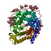
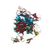
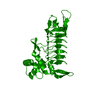
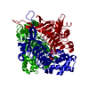
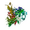
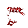
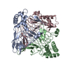
| Title | Crystal structure of death receptor 4 (DR4; TNFFRSF10A) bound to TRAIL (TNFSF10) | ||||||
 Components Components |
| ||||||
 Keywords Keywords | APOPTOSIS / binding and specificity / ligand-receptor complex / TNF receptor family | ||||||
| Function / homology |  Function and homology information Function and homology informationTRAIL binding / death receptor activity / TRAIL signaling / TRAIL-activated apoptotic signaling pathway / Regulation by c-FLIP / CASP8 activity is inhibited / Dimerization of procaspase-8 / Caspase activation via Death Receptors in the presence of ligand / activation of NF-kappaB-inducing kinase activity / tumor necrosis factor receptor binding ...TRAIL binding / death receptor activity / TRAIL signaling / TRAIL-activated apoptotic signaling pathway / Regulation by c-FLIP / CASP8 activity is inhibited / Dimerization of procaspase-8 / Caspase activation via Death Receptors in the presence of ligand / activation of NF-kappaB-inducing kinase activity / tumor necrosis factor receptor binding / TP53 Regulates Transcription of Death Receptors and Ligands / positive regulation of extrinsic apoptotic signaling pathway / RIPK1-mediated regulated necrosis / positive regulation of release of cytochrome c from mitochondria / extrinsic apoptotic signaling pathway via death domain receptors / plasma membrane raft / extrinsic apoptotic signaling pathway / cytokine activity / Cell surface interactions at the vascular wall / cellular response to mechanical stimulus / cell-cell signaling / signaling receptor activity / protease binding / positive regulation of canonical NF-kappaB signal transduction / cell surface receptor signaling pathway / immune response / positive regulation of apoptotic process / membrane raft / signaling receptor binding / apoptotic process / Golgi apparatus / cell surface / signal transduction / : / extracellular exosome / extracellular region / zinc ion binding / identical protein binding / plasma membrane / cytosol Similarity search - Function | ||||||
| Biological species |  Homo sapiens (human) Homo sapiens (human) | ||||||
| Method |  X-RAY DIFFRACTION / SYNCHROTRON / MOLECULAR REPLACEMENT / Resolution: 3 Å X-RAY DIFFRACTION / SYNCHROTRON / MOLECULAR REPLACEMENT / Resolution: 3 Å | ||||||
 Authors Authors | Sheriff, S. | ||||||
 Citation Citation | Journal: Acta Crystallogr F Struct Biol Commun / Year: 2015 Title: The structure of the death receptor 4-TNF-related apoptosis-inducing ligand (DR4-TRAIL) complex. Authors: Ramamurthy, V. / Yamniuk, A.P. / Lawrence, E.J. / Yong, W. / Schneeweis, L.A. / Cheng, L. / Murdock, M. / Corbett, M.J. / Doyle, M.L. / Sheriff, S. | ||||||
| History |
|
- Structure visualization
| Structure viewer | Molecule: MolmilJmol/JSmol |
|---|
- Downloads & links
-Download
| PDBx/mmCIF format | 5cir.cif.gz | 156.3 KB | Display | PDBx/mmCIF format |
|---|---|---|---|---|
| PDB format | pdb5cir.ent.gz | 119.6 KB | Display | PDB format |
| PDBx/mmJSON format | 5cir.json.gz | Tree view | PDBx/mmJSON format | |
| Others |  Other downloads Other downloads |
-Validation report
| Arichive directory | https://data.pdbj.org/pub/pdb/validation_reports/ci/5cirftp://data.pdbj.org/pub/pdb/validation_reports/ci/5cir | HTTPS FTP |
|---|
-Related structure data


-Links
 PDBj
PDBj


- Assembly
| Deposited unit | 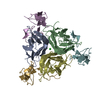
| ||||||||||||||||||||||||||||||
|---|---|---|---|---|---|---|---|---|---|---|---|---|---|---|---|---|---|---|---|---|---|---|---|---|---|---|---|---|---|---|---|
| 1 |
| ||||||||||||||||||||||||||||||
| Unit cell |
| ||||||||||||||||||||||||||||||
| Noncrystallographic symmetry (NCS) | NCS oper:
|
-Components
| #1: Protein | Mass: 19652.049 Da / Num. of mol.: 3 / Fragment: Residues 114-281 Source method: isolated from a genetically manipulated source Source: (gene. exp.) Homo sapiens (human) / Gene: TNFSF10, APO2L, TRAIL / Plasmid: pET28-NHis-TEV / Production host:  #2: Protein | Mass: 11749.284 Da / Num. of mol.: 3 / Fragment: Extracellular domain residues 125-232 Source method: isolated from a genetically manipulated source Source: (gene. exp.) Homo sapiens (human) / Gene: TNFRSF10A, APO2, DR4, TRAILR1 / Plasmid: pET28 / Production host: #3: Chemical | ChemComp-ZN / |   Mass: 65.409 Da / Num. of mol.: 1 / Source method: obtained synthetically / Formula: Zn Mass: 65.409 Da / Num. of mol.: 1 / Source method: obtained synthetically / Formula: Zn#4: Chemical | ChemComp-CL / |   Mass: 35.453 Da / Num. of mol.: 1 / Source method: obtained synthetically / Formula: Cl Mass: 35.453 Da / Num. of mol.: 1 / Source method: obtained synthetically / Formula: Cl#5: Water | ChemComp-HOH / |  Mass: 18.015 Da / Num. of mol.: 8 / Source method: isolated from a natural source / Formula: H2O Mass: 18.015 Da / Num. of mol.: 8 / Source method: isolated from a natural source / Formula: H2OHas protein modification | Y | |
|---|
-Experimental details
-Experiment
| Experiment | Method: X-RAY DIFFRACTION / Number of used crystals: 1 |
|---|
- Sample preparation
| Crystal | Density Matthews: 2.12 Å3/Da / Density % sol: 41.93 % |
|---|---|
| Crystal grow | Temperature: 293 K / Method: vapor diffusion, hanging drop / pH: 5 Details: 100 mM MMT (1:2:2 ratio of DL-malic acid, MES, Tris base), pH 5.0, 22.2%(W/V) PEG 2000MME |
-Data collection
| Diffraction | Mean temperature: 100 K |
|---|---|
| Diffraction source | Source: SYNCHROTRON / Site: APS  / Beamline: 17-ID / Wavelength: 1 Å / Beamline: 17-ID / Wavelength: 1 Å |
| Detector | Type: MAR CCD 165 mm / Detector: CCD / Date: Jul 6, 2009 |
| Radiation | Protocol: SINGLE WAVELENGTH / Monochromatic (M) / Laue (L): M / Scattering type: x-ray |
| Radiation wavelength | Wavelength: 1 Å / Relative weight: 1 |
| Reflection | Resolution: 3→50 Å / Num. obs: 16742 / % possible obs: 99.8 % / Observed criterion σ(I): 0 / Redundancy: 7.3 % / Biso Wilson estimate: 71.79 Å2 / Rmerge(I) obs: 0.092 / Net I/σ(I): 20.8 |
| Reflection shell | Resolution: 3→3.11 Å / Redundancy: 7.5 % / Rmerge(I) obs: 0.365 / Mean I/σ(I) obs: 6.4 / Rejects: 0 / % possible all: 100 |
- Processing
| Software |
| ||||||||||||||||||||||||||||||||||||||||||||||||||||||||||||||||||||||||||||||||||||||||||||||||||||||||||||
|---|---|---|---|---|---|---|---|---|---|---|---|---|---|---|---|---|---|---|---|---|---|---|---|---|---|---|---|---|---|---|---|---|---|---|---|---|---|---|---|---|---|---|---|---|---|---|---|---|---|---|---|---|---|---|---|---|---|---|---|---|---|---|---|---|---|---|---|---|---|---|---|---|---|---|---|---|---|---|---|---|---|---|---|---|---|---|---|---|---|---|---|---|---|---|---|---|---|---|---|---|---|---|---|---|---|---|---|---|---|
| Refinement | Method to determine structure: MOLECULAR REPLACEMENT Starting model: TRAIL FROM 1D0G AND DR4 FROM 1DOG DR5 Resolution: 3→27.6 Å / Cor.coef. Fo:Fc: 0.9265 / Cor.coef. Fo:Fc free: 0.8932 / Cross valid method: THROUGHOUT / σ(F): 0 / SU Rfree Blow DPI: 0.372 Details: Non-crystallographic symmetry was used in the form of Local Structure Similarity Restraints (LSSR) with automatic pruning of discrepant residues and with a TARGET_WEIGHT OF 0.5.
| ||||||||||||||||||||||||||||||||||||||||||||||||||||||||||||||||||||||||||||||||||||||||||||||||||||||||||||
| Displacement parameters | Biso max: 135.15 Å2 / Biso mean: 42.22 Å2 / Biso min: 7.15 Å2
| ||||||||||||||||||||||||||||||||||||||||||||||||||||||||||||||||||||||||||||||||||||||||||||||||||||||||||||
| Refine analyze | Luzzati coordinate error obs: 0.309 Å | ||||||||||||||||||||||||||||||||||||||||||||||||||||||||||||||||||||||||||||||||||||||||||||||||||||||||||||
| Refinement step | Cycle: final / Resolution: 3→27.6 Å
| ||||||||||||||||||||||||||||||||||||||||||||||||||||||||||||||||||||||||||||||||||||||||||||||||||||||||||||
| Refine LS restraints |
| ||||||||||||||||||||||||||||||||||||||||||||||||||||||||||||||||||||||||||||||||||||||||||||||||||||||||||||
| LS refinement shell | Resolution: 3→3.21 Å / Total num. of bins used: 8
|
