 ムービー
ムービー コントローラー


+ データを開く

- 基本情報
基本情報
| 登録情報 | データベース: PDB / ID: 5c9v | |||||||||
|---|---|---|---|---|---|---|---|---|---|---|
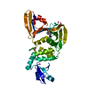
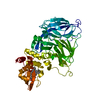
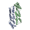
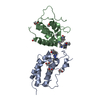
| タイトル | Structure of human Parkin G319A | |||||||||
 要素 要素 | E3 ubiquitin-protein ligase parkin | |||||||||
 キーワード キーワード | SIGNALING PROTEIN / Parkin / ubiquitin / E3 ligase / RBR / Parkinson's disease / mitophagy / cell signalling | |||||||||
| 機能・相同性 |  機能・相同性情報 機能・相同性情報: / positive regulation of retrograde transport, endosome to Golgi / regulation of lipid transport / positive regulation of neurotransmitter uptake / negative regulation of endoplasmic reticulum stress-induced neuron intrinsic apoptotic signaling pathway / negative regulation of spontaneous neurotransmitter secretion / negative regulation of intralumenal vesicle formation / regulation protein catabolic process at presynapse / cellular response to L-glutamine / regulation of protein targeting to mitochondrion ...: / positive regulation of retrograde transport, endosome to Golgi / regulation of lipid transport / positive regulation of neurotransmitter uptake / negative regulation of endoplasmic reticulum stress-induced neuron intrinsic apoptotic signaling pathway / negative regulation of spontaneous neurotransmitter secretion / negative regulation of intralumenal vesicle formation / regulation protein catabolic process at presynapse / cellular response to L-glutamine / regulation of protein targeting to mitochondrion / negative regulation of exosomal secretion / negative regulation of glucokinase activity / mitochondrion to lysosome vesicle-mediated transport / type 2 mitophagy / response to curcumin / negative regulation of mitochondrial fusion / cellular response to hydrogen sulfide / Parkin-FBXW7-Cul1 ubiquitin ligase complex / protein K29-linked ubiquitination / free ubiquitin chain polymerization / Lewy body / positive regulation of protein linear polyubiquitination / host-mediated suppression of viral genome replication / protein K27-linked ubiquitination / RBR-type E3 ubiquitin transferase / regulation of synaptic vesicle transport / positive regulation of mitophagy / F-box domain binding / positive regulation of mitochondrial fusion / negative regulation of actin filament bundle assembly / regulation of necroptotic process / mitochondrial fragmentation involved in apoptotic process / regulation of cellular response to oxidative stress / positive regulation of dendrite extension / regulation of dopamine metabolic process / negative regulation of excitatory postsynaptic potential / protein K6-linked ubiquitination / norepinephrine metabolic process / dopaminergic synapse / autophagy of mitochondrion / positive regulation of type 2 mitophagy / mitochondrion localization / protein localization to mitochondrion / cellular response to dopamine / positive regulation of proteasomal protein catabolic process / positive regulation of protein localization to membrane / cellular response to toxic substance / mitochondrial fission / positive regulation of tumor necrosis factor-mediated signaling pathway / protein K11-linked ubiquitination / negative regulation of intrinsic apoptotic signaling pathway by p53 class mediator / negative regulation of oxidative stress-induced neuron intrinsic apoptotic signaling pathway / aggresome assembly / cellular response to L-glutamate / regulation of mitochondrion organization / negative regulation of synaptic transmission, glutamatergic / ubiquitin conjugating enzyme binding / regulation of canonical Wnt signaling pathway / negative regulation of JNK cascade / aggresome / positive regulation of mitochondrial membrane potential / regulation of reactive oxygen species metabolic process / response to corticosterone / positive regulation of mitochondrial fission / response to muscle activity / negative regulation of release of cytochrome c from mitochondria / ubiquitin-specific protease binding / startle response / dopamine metabolic process / dopamine uptake involved in synaptic transmission / regulation of dopamine secretion / positive regulation of ATP biosynthetic process / negative regulation of reactive oxygen species metabolic process / cullin family protein binding / regulation of glucose metabolic process / regulation of protein ubiquitination / protein K63-linked ubiquitination / protein deubiquitination / protein monoubiquitination / cellular response to unfolded protein / ubiquitin ligase complex / regulation of synaptic vesicle endocytosis / negative regulation of mitochondrial fission / positive regulation of insulin secretion involved in cellular response to glucose stimulus / regulation of postsynaptic membrane neurotransmitter receptor levels / protein K48-linked ubiquitination / negative regulation of endoplasmic reticulum stress-induced intrinsic apoptotic signaling pathway / protein autoubiquitination / mitophagy / proteasomal protein catabolic process / phospholipase binding / negative regulation of reactive oxygen species biosynthetic process / cellular response to manganese ion / heat shock protein binding / ERAD pathway / Hsp70 protein binding / tubulin binding / response to endoplasmic reticulum stress / Josephin domain DUBs / central nervous system development 類似検索 - 分子機能 | |||||||||
| 生物種 |  Homo sapiens (ヒト) Homo sapiens (ヒト) | |||||||||
| 手法 |  X線回折 / シンクロトロン / 分子置換 / 解像度: 2.35 Å X線回折 / シンクロトロン / 分子置換 / 解像度: 2.35 Å | |||||||||
 データ登録者 データ登録者 | Wauer, T. / Komander, D. | |||||||||
| 資金援助 |  英国, 2件 英国, 2件
| |||||||||
 引用 引用 | ジャーナル: Nature / 年: 2015 タイトル: Mechanism of phospho-ubiquitin-induced PARKIN activation. 著者: Wauer, T. / Simicek, M. / Schubert, A. / Komander, D. #1: ジャーナル: EMBO J. / 年: 2013タイトル: Structure of the human Parkin ligase domain in an autoinhibited state. 著者: Wauer, T. / Komander, D. | |||||||||
| 履歴 |
|
- 構造の表示
構造の表示
| 構造ビューア | 分子: MolmilJmol/JSmol |
|---|
- ダウンロードとリンク
-ダウンロード
| PDBx/mmCIF形式 | 5c9v.cif.gz | 138.6 KB | 表示 | PDBx/mmCIF形式 |
|---|---|---|---|---|
| PDB形式 | pdb5c9v.ent.gz | 107 KB | 表示 | PDB形式 |
| PDBx/mmJSON形式 | 5c9v.json.gz | ツリー表示 | PDBx/mmJSON形式 | |
| その他 |  その他のダウンロード その他のダウンロード |
-検証レポート
| アーカイブディレクトリ | https://data.pdbj.org/pub/pdb/validation_reports/c9/5c9vftp://data.pdbj.org/pub/pdb/validation_reports/c9/5c9v | HTTPS FTP |
|---|
-関連構造データ


-リンク
 PDBj
PDBj

- 集合体
集合体
| 登録構造単位 | 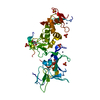
| |||||||||||||||
|---|---|---|---|---|---|---|---|---|---|---|---|---|---|---|---|---|
| 1 |
| |||||||||||||||
| 単位格子 |
| |||||||||||||||
| Components on special symmetry positions |
|
-要素
| #1: タンパク質 | 分子量: 36892.129 Da / 分子数: 1 / 断片: UNP residues 137-465 / 変異: G319A / 由来タイプ: 組換発現 / 詳細: engineered mutation at position G319A / 由来: (組換発現) Homo sapiens (ヒト) / 遺伝子: PARK2, PRKN / プラスミド: pOPINK発現宿主:  参照: UniProt: O60260, 合成酵素; C-N結合を形成; 酸-D-アミノ酸リガーゼ(ペプチド合成) | ||||||
|---|---|---|---|---|---|---|---|
| #2: 化合物 | ChemComp-ZN /   分子量: 65.409 Da / 分子数: 8 / 由来タイプ: 合成 / 式: Zn 分子量: 65.409 Da / 分子数: 8 / 由来タイプ: 合成 / 式: Zn#3: 化合物 | ChemComp-SO4 /   分子量: 96.063 Da / 分子数: 6 / 由来タイプ: 合成 / 式: SO4 分子量: 96.063 Da / 分子数: 6 / 由来タイプ: 合成 / 式: SO4#4: 化合物 |   分子量: 92.094 Da / 分子数: 3 / 由来タイプ: 合成 / 式: C3H8O3 分子量: 92.094 Da / 分子数: 3 / 由来タイプ: 合成 / 式: C3H8O3#5: 水 | ChemComp-HOH / |  分子量: 18.015 Da / 分子数: 81 / 由来タイプ: 天然 / 式: H2O 分子量: 18.015 Da / 分子数: 81 / 由来タイプ: 天然 / 式: H2O |
-実験情報
-実験
| 実験 | 手法: X線回折 |
|---|
- 試料調製
試料調製
| 結晶 | マシュー密度: 3.63 Å3/Da / 溶媒含有率: 66.1 % |
|---|---|
| 結晶化 | 温度: 291 K / 手法: 蒸気拡散法, シッティングドロップ法 / pH: 5.6 詳細: 1.8 M lithium sulphate, 0.01 M MgCl2, 0.05 M MES pH 5.6 PH範囲: 5.6 |
-データ収集
| 回折 | 平均測定温度: 100 K |
|---|---|
| 放射光源 | 由来: シンクロトロン / サイト: Diamond / ビームライン: I04-1 / 波長: 0.9173 Å |
| 検出器 | タイプ: DECTRIS PILATUS 6M-F / 検出器: PIXEL / 日付: 2015年4月20日 |
| 放射 | プロトコル: SINGLE WAVELENGTH / 単色(M)・ラウエ(L): M / 散乱光タイプ: x-ray |
| 放射波長 | 波長: 0.9173 Å / 相対比: 1 |
| 反射 | 解像度: 2.35→86.68 Å / Num. obs: 22270 / % possible obs: 100 % / Observed criterion σ(I): 2 / 冗長度: 6.9 % / Biso Wilson estimate: 42.5 Å2 / Rmerge(I) obs: 0.089 / Net I/σ(I): 13.3 |
| 反射 シェル | 解像度: 2.35→2.43 Å / 冗長度: 6.8 % / Rmerge(I) obs: 0.8 / Mean I/σ(I) obs: 2.1 / % possible all: 100 |
- 解析
解析
| ソフトウェア |
| |||||||||||||||||||||||||||||||||||||||||||||||||||||||||||||||
|---|---|---|---|---|---|---|---|---|---|---|---|---|---|---|---|---|---|---|---|---|---|---|---|---|---|---|---|---|---|---|---|---|---|---|---|---|---|---|---|---|---|---|---|---|---|---|---|---|---|---|---|---|---|---|---|---|---|---|---|---|---|---|---|---|
| 精密化 | 構造決定の手法: 分子置換 開始モデル: 4bm9 解像度: 2.35→84.68 Å / SU ML: 0.22 / 交差検証法: FREE R-VALUE / σ(F): 1.33 / 位相誤差: 23.8 / 立体化学のターゲット値: ML
| |||||||||||||||||||||||||||||||||||||||||||||||||||||||||||||||
| 溶媒の処理 | 減衰半径: 0.9 Å / VDWプローブ半径: 1.11 Å / 溶媒モデル: FLAT BULK SOLVENT MODEL | |||||||||||||||||||||||||||||||||||||||||||||||||||||||||||||||
| 原子変位パラメータ | Biso mean: 54.3 Å2 | |||||||||||||||||||||||||||||||||||||||||||||||||||||||||||||||
| 精密化ステップ | サイクル: LAST / 解像度: 2.35→84.68 Å
| |||||||||||||||||||||||||||||||||||||||||||||||||||||||||||||||
| 拘束条件 |
| |||||||||||||||||||||||||||||||||||||||||||||||||||||||||||||||
| LS精密化 シェル |
| |||||||||||||||||||||||||||||||||||||||||||||||||||||||||||||||
| 精密化 TLS | 手法: refined / Origin x: 7.8845 Å / Origin y: 31.4194 Å / Origin z: 36.9402 Å
| |||||||||||||||||||||||||||||||||||||||||||||||||||||||||||||||
| 精密化 TLSグループ | Selection details: chain A |
