Resolution: 2.4→2.44 Å / Redundancy: 3.4 % / Rmerge(I) obs: 0.419 / Mean I/σ(I) obs: 2.67 / % possible all: 33.6
-
Processing
Software
Name
Version
Classification
REFMAC
5.7.0032
refinement
HKL-2000
datareduction
SCALEPACK
datascaling
SOLVE
phasing
Refinement
Resolution: 2.4→47.37 Å / Cor.coef. Fo:Fc: 0.953 / Cor.coef. Fo:Fc free: 0.917 / SU B: 16.365 / SU ML: 0.197 / Cross valid method: THROUGHOUT / ESU R: 0.296 / ESU R Free: 0.25 / Stereochemistry target values: MAXIMUM LIKELIHOOD / Details: HYDROGENS HAVE BEEN ADDED IN THE RIDING POSITIONS
Rfactor
Num. reflection
% reflection
Selection details
Rfree
0.26222
1458
5.1 %
RANDOM
Rwork
0.19893
-
-
-
obs
0.20206
27301
91.02 %
-
Solvent computation
Ion probe radii: 0.8 Å / Shrinkage radii: 0.8 Å / VDW probe radii: 1.2 Å / Solvent model: MASK
 Movie
Movie Controller

 Yorodumi
 Open data
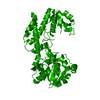
 Basic information
 Components
 Keywords
 Function and homology information
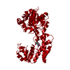
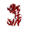
 Rickettsia conorii (bacteria)
 X-RAY DIFFRACTION /
  Authors
 Citation
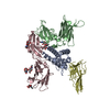
 Structure visualization
 Downloads & links
 Other downloads


 PDBj
PDBj
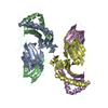
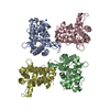
 Assembly
 Mass: 18.015 Da / Num. of mol.: 128 / Source method: isolated from a natural source / Formula: H2O
 Sample preparation
 / Beamline: 22-ID / Wavelength: 0.9825 Å
 Processing