 Movie
Movie Controller

[English] 日本語
 Yorodumi
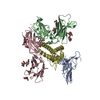
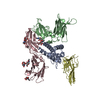
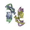
- PDB-3plr: Crystal structure of Klebsiella pneumoniae UDP-glucose 6-dehydrog... -
+ Open data

- Basic information
| Entry | Database: PDB / ID: 3plr | ||||||
|---|---|---|---|---|---|---|---|
| Title | Crystal structure of Klebsiella pneumoniae UDP-glucose 6-dehydrogenase complexed with NADH and UDP-glucose | ||||||
 Components Components | UDP-glucose 6-dehydrogenase | ||||||
 Keywords Keywords | OXIDOREDUCTASE / Rossmann Fold / Dehydrogenase | ||||||
| Function / homology |  Function and homology information Function and homology informationUDP-glucose 6-dehydrogenase / UDP-glucose 6-dehydrogenase activity / UDP-glucuronate biosynthetic process / polysaccharide biosynthetic process / NAD binding Similarity search - Function | ||||||
| Biological species |  Klebsiella pneumoniae (bacteria) Klebsiella pneumoniae (bacteria) | ||||||
| Method |  X-RAY DIFFRACTION / SYNCHROTRON / MOLECULAR REPLACEMENT / Resolution: 1.7 Å X-RAY DIFFRACTION / SYNCHROTRON / MOLECULAR REPLACEMENT / Resolution: 1.7 Å | ||||||
 Authors Authors | Chen, Y.-Y. / Ko, T.-P. / Lin, C.-H. / Chen, W.-H. / Wang, A.H.-J. | ||||||
 Citation Citation | Journal: J.Struct.Biol. / Year: 2011 Title: Conformational change upon product binding to Klebsiella pneumoniae UDP-glucose dehydrogenase: a possible inhibition mechanism for the key enzyme in polymyxin resistance. Authors: Chen, Y.Y. / Ko, T.P. / Lin, C.H. / Chen, W.H. / Wang, A.H. | ||||||
| History |
|
- Structure visualization
| Structure viewer | Molecule: MolmilJmol/JSmol |
|---|
- Downloads & links
-Download
| PDBx/mmCIF format | 3plr.cif.gz | 101.5 KB | Display | PDBx/mmCIF format |
|---|---|---|---|---|
| PDB format | pdb3plr.ent.gz | 74.6 KB | Display | PDB format |
| PDBx/mmJSON format | 3plr.json.gz | Tree view | PDBx/mmJSON format | |
| Others |  Other downloads Other downloads |
-Validation report
| Arichive directory | https://data.pdbj.org/pub/pdb/validation_reports/pl/3plrftp://data.pdbj.org/pub/pdb/validation_reports/pl/3plr | HTTPS FTP |
|---|
-Related structure data
| Related structure data | 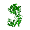 3phlC 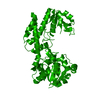 3pidC 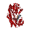 3pjgC 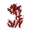 3plnSC S: Starting model for refinement C: citing same article ( |
|---|---|
| Similar structure data |


-Links
 PDBj
PDBj


- Assembly
| Deposited unit | 
| |||||||||
|---|---|---|---|---|---|---|---|---|---|---|
| 1 | 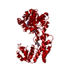
| |||||||||
| Unit cell |
| |||||||||
| Components on special symmetry positions |
|
-Components
| #1: Protein | Mass: 47385.664 Da / Num. of mol.: 1 Source method: isolated from a genetically manipulated source Source: (gene. exp.) Klebsiella pneumoniae (bacteria) / Strain: NTUH-K2044 / Gene: KP1_3701, ugd / Plasmid: pET28a / Production host: References: UniProt: C4XAX5, UniProt: A0A0J9WZA6*PLUS, UDP-glucose 6-dehydrogenase |
|---|---|
| #2: Chemical | ChemComp-NAI /   Mass: 665.441 Da / Num. of mol.: 1 / Source method: obtained synthetically / Formula: C21H29N7O14P2 Mass: 665.441 Da / Num. of mol.: 1 / Source method: obtained synthetically / Formula: C21H29N7O14P2 |
| #3: Chemical | ChemComp-U5P /   Mass: 324.181 Da / Num. of mol.: 1 / Source method: obtained synthetically / Formula: C9H13N2O9P Mass: 324.181 Da / Num. of mol.: 1 / Source method: obtained synthetically / Formula: C9H13N2O9P |
| #4: Water | ChemComp-HOH /  Mass: 18.015 Da / Num. of mol.: 428 / Source method: isolated from a natural source / Formula: H2O Mass: 18.015 Da / Num. of mol.: 428 / Source method: isolated from a natural source / Formula: H2O |
-Experimental details
-Experiment
| Experiment | Method: X-RAY DIFFRACTION / Number of used crystals: 1 |
|---|
- Sample preparation
| Crystal | Density Matthews: 2.6 Å3/Da / Density % sol: 52.66 % |
|---|---|
| Crystal grow | Temperature: 283 K / Method: vapor diffusion, sitting drop / pH: 8 Details: 0.1M Tris-HCl buffer (pH 8.0), 0.8M sodium formate, 19-25 % (w/v) PEG 2000 monomethyl ether, 2mM UPG, 5mM NADH, VAPOR DIFFUSION, SITTING DROP, temperature 283K |
-Data collection
| Diffraction | Mean temperature: 100 K |
|---|---|
| Diffraction source | Source: SYNCHROTRON / Site: NSRRC  / Beamline: BL13B1 / Wavelength: 1 Å / Beamline: BL13B1 / Wavelength: 1 Å |
| Detector | Type: ADSC QUANTUM 315 / Detector: CCD / Date: May 15, 2009 / Details: mirrors |
| Radiation | Monochromator: SAGITALLY FOCUSED Si(111) / Protocol: SINGLE WAVELENGTH / Monochromatic (M) / Laue (L): M / Scattering type: x-ray |
| Radiation wavelength | Wavelength: 1 Å / Relative weight: 1 |
| Reflection | Resolution: 1.7→30 Å / Num. all: 53574 / Num. obs: 53413 / % possible obs: 99.7 % / Redundancy: 5.7 % / Rmerge(I) obs: 0.063 / Net I/σ(I): 26.5 |
| Reflection shell | Resolution: 1.7→1.76 Å / Redundancy: 5.6 % / Rmerge(I) obs: 0.456 / Mean I/σ(I) obs: 3.4 / Num. unique all: 5286 / % possible all: 99.9 |
- Processing
| Software |
| |||||||||||||||||||||||||
|---|---|---|---|---|---|---|---|---|---|---|---|---|---|---|---|---|---|---|---|---|---|---|---|---|---|---|
| Refinement | Method to determine structure: MOLECULAR REPLACEMENT Starting model: 3PLN Resolution: 1.7→30 Å / Cross valid method: THROUGHOUT / σ(F): 0 / σ(I): 0 / Stereochemistry target values: Engh & Huber
| |||||||||||||||||||||||||
| Refine analyze |
| |||||||||||||||||||||||||
| Refinement step | Cycle: LAST / Resolution: 1.7→30 Å
| |||||||||||||||||||||||||
| Refine LS restraints |
| |||||||||||||||||||||||||
| LS refinement shell | Resolution: 1.7→1.76 Å / Rfactor Rfree error: 0.0012
|
