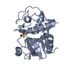
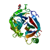
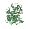
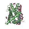
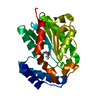
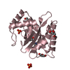
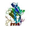
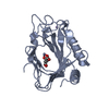
- PDB-5c5t: The crystal structure of viral collagen prolyl hydroxylase vCPH f... -
+
Open data
ID or keywords:
Loading...
-
Basic information
Entry
Database: PDB / ID: 5c5t
Title
The crystal structure of viral collagen prolyl hydroxylase vCPH from Paramecium Bursaria Chlorella virus-1 - 2OG complex
Components
Prolyl 4-hydroxylase
Keywords
OXIDOREDUCTASE / dioxygenase / prolyl hydroxylase
Function / homology
Function and homology information
L-ascorbic acid binding / dioxygenase activity / oxidoreductase activity, acting on paired donors, with incorporation or reduction of molecular oxygen / iron ion binding Similarity search - Function
Mass: 18.015 Da / Num. of mol.: 396 / Source method: isolated from a natural source / Formula: H2O
Has protein modification
Y
-
Experimental details
-
Experiment
Experiment
Method: X-RAY DIFFRACTION
-
Sample preparation
Crystal
Density Matthews: 2.11 Å3/Da / Density % sol: 41.81 %
Crystal grow
Temperature: 277 K / Method: vapor diffusion, sitting drop / pH: 7.5 Details: 0.1 M [Imidazole; MES monohydrate (acid)], 0.1 M amino acids [0.2 M L-Na-Glutamate; 0.2 M Alanine (racemic); 0.2 M Glycine; 0.2M Lysine HCL (racemic); 0.2 M Serine (racemic)], 30 % PEGMME ...Details: 0.1 M [Imidazole; MES monohydrate (acid)], 0.1 M amino acids [0.2 M L-Na-Glutamate; 0.2 M Alanine (racemic); 0.2 M Glycine; 0.2M Lysine HCL (racemic); 0.2 M Serine (racemic)], 30 % PEGMME 550 PEG 20K pH 6.5 [Morpheus HT96 condition H1, Molecular Dimensions] COmplex generated by soaking crystals in mother liquor supplemented with 250 mM ZnSO4 and 100 mM 2-OG for a period of 16 hours.
In the structure databanks used in Yorodumi, some data are registered as the other names, "COVID-19 virus" and "2019-nCoV". Here are the details of the virus and the list of structure data.
Jan 31, 2019. EMDB accession codes are about to change! (news from PDBe EMDB page)
EMDB accession codes are about to change! (news from PDBe EMDB page)
The allocation of 4 digits for EMDB accession codes will soon come to an end. Whilst these codes will remain in use, new EMDB accession codes will include an additional digit and will expand incrementally as the available range of codes is exhausted. The current 4-digit format prefixed with “EMD-” (i.e. EMD-XXXX) will advance to a 5-digit format (i.e. EMD-XXXXX), and so on. It is currently estimated that the 4-digit codes will be depleted around Spring 2019, at which point the 5-digit format will come into force.
The EM Navigator/Yorodumi systems omit the EMD- prefix.
Related info.:Q: What is EMD? / ID/Accession-code notation in Yorodumi/EM Navigator
Yorodumi is a browser for structure data from EMDB, PDB, SASBDB, etc.
This page is also the successor to EM Navigator detail page, and also detail information page/front-end page for Omokage search.
The word "yorodu" (or yorozu) is an old Japanese word meaning "ten thousand". "mi" (miru) is to see.
Related info.:EMDB / PDB / SASBDB / Comparison of 3 databanks / Yorodumi Search / Aug 31, 2016. New EM Navigator & Yorodumi / Yorodumi Papers / Jmol/JSmol / Function and homology information / Changes in new EM Navigator and Yorodumi
 Movie
Movie Controller

 Yorodumi
 Open data
 Basic information
 Components
 Keywords
 Function and homology information
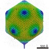 Paramecium bursaria Chlorella virus 1
 X-RAY DIFFRACTION /
  Authors
 United Kingdom, 1items
  Citation
 Structure visualization
 Downloads & links
 Other downloads


 PDBj
PDBj


 Assembly


 Mass: 146.098 Da / Num. of mol.: 2 / Source method: obtained synthetically / Formula: C5H6O5
 Mass: 65.409 Da / Num. of mol.: 2 / Source method: obtained synthetically / Formula: Zn
 Mass: 54.938 Da / Num. of mol.: 1 / Source method: obtained synthetically / Formula: Mn
 Mass: 18.015 Da / Num. of mol.: 396 / Source method: isolated from a natural source / Formula: H2O
 Sample preparation
 Processing