 Movie
Movie Controller

[English] 日本語
 Yorodumi
- PDB-1o0e: 1.9 Angstrom Crystal Structure of a plant cysteine protease Ervat... -
+ Open data

- Basic information
| Entry | Database: PDB / ID: 1o0e | ||||||
|---|---|---|---|---|---|---|---|
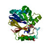
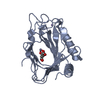
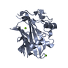
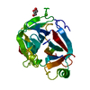
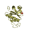
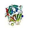
| Title | 1.9 Angstrom Crystal Structure of a plant cysteine protease Ervatamin C | ||||||
 Components Components | Ervatamin C | ||||||
 Keywords Keywords | HYDROLASE / Plant cysteine protease / two domain / stable at pH 2-12 | ||||||
| Function / homology |  Function and homology information Function and homology informationHydrolases; Acting on peptide bonds (peptidases); Cysteine endopeptidases / cysteine-type endopeptidase activity / proteolysis / extracellular region Similarity search - Function | ||||||
| Biological species |  Tabernaemontana divaricata (pinwheelflower) Tabernaemontana divaricata (pinwheelflower) | ||||||
| Method |  X-RAY DIFFRACTION / MOLECULAR REPLACEMENT / Resolution: 1.9 Å X-RAY DIFFRACTION / MOLECULAR REPLACEMENT / Resolution: 1.9 Å | ||||||
 Authors Authors | Thakurta, P.G. / Chakrabarti, C. / Biswas, S. / Dattagupta, J.K. | ||||||
 Citation Citation | Journal: Biochemistry / Year: 2004 Title: Structural Basis of the Unusual Stability and Substrate Specificity of Ervatamin C, a Plant Cysteine Protease from Ervatamia coronaria Authors: Thakurta, P.G. / Biswas, S. / Chakrabarti, C. / Sundd, M. / Jagannadham, M.V. / Dattagupta, J.K. | ||||||
| History |
|
- Structure visualization
| Structure viewer | Molecule: MolmilJmol/JSmol |
|---|
- Downloads & links
-Download
| PDBx/mmCIF format | 1o0e.cif.gz | 93.8 KB | Display | PDBx/mmCIF format |
|---|---|---|---|---|
| PDB format | pdb1o0e.ent.gz | 72.3 KB | Display | PDB format |
| PDBx/mmJSON format | 1o0e.json.gz | Tree view | PDBx/mmJSON format | |
| Others |  Other downloads Other downloads |
-Validation report
| Arichive directory | https://data.pdbj.org/pub/pdb/validation_reports/o0/1o0eftp://data.pdbj.org/pub/pdb/validation_reports/o0/1o0e | HTTPS FTP |
|---|
-Related structure data
| Related structure data |  1iwdS S: Starting model for refinement |
|---|---|
| Similar structure data |


-Links
 PDBj
PDBj
- Assembly
| Deposited unit | 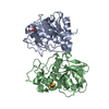
| ||||||||
|---|---|---|---|---|---|---|---|---|---|
| 1 | 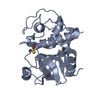
| ||||||||
| 2 | 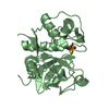
| ||||||||
| Unit cell |
|
-Components
| #1: Protein | Mass: 22516.463 Da / Num. of mol.: 2 / Source method: isolated from a natural source Source: (natural) Tabernaemontana divaricata (pinwheelflower)References: UniProt: P83654 #2: Chemical |   Mass: 112.128 Da / Num. of mol.: 2 / Source method: obtained synthetically / Formula: O3S2 Mass: 112.128 Da / Num. of mol.: 2 / Source method: obtained synthetically / Formula: O3S2#3: Water | ChemComp-HOH / |  Mass: 18.015 Da / Num. of mol.: 256 / Source method: isolated from a natural source / Formula: H2O Mass: 18.015 Da / Num. of mol.: 256 / Source method: isolated from a natural source / Formula: H2OHas protein modification | Y | |
|---|
-Experimental details
-Experiment
| Experiment | Method: X-RAY DIFFRACTION / Number of used crystals: 1 |
|---|
- Sample preparation
| Crystal | Density Matthews: 2.51 Å3/Da / Density % sol: 50.68 % | ||||||||||||||||||||||||||||||
|---|---|---|---|---|---|---|---|---|---|---|---|---|---|---|---|---|---|---|---|---|---|---|---|---|---|---|---|---|---|---|---|
| Crystal grow | Temperature: 293 K / Method: vapor diffusion, hanging drop / pH: 7 Details: PEG8000, potassium phosphate monobasic, pH 7.0, VAPOR DIFFUSION, HANGING DROP, temperature 293K | ||||||||||||||||||||||||||||||
| Crystal grow | *PLUS Method: vapor diffusion, hanging drop | ||||||||||||||||||||||||||||||
| Components of the solutions | *PLUS
|
-Data collection
| Diffraction | Mean temperature: 293 K |
|---|---|
| Diffraction source | Source: ROTATING ANODE / Type: RIGAKU RU200B / Wavelength: 1.5418 Å |
| Detector | Type: MARRESEARCH / Detector: IMAGE PLATE / Date: Jan 4, 1999 / Details: OSMIC MAXFLUX CONFOCAL OPTICS |
| Radiation | Protocol: SINGLE WAVELENGTH / Monochromatic (M) / Laue (L): M / Scattering type: x-ray |
| Radiation wavelength | Wavelength: 1.5418 Å / Relative weight: 1 |
| Reflection | Resolution: 1.9→15 Å / Num. all: 38978 / Num. obs: 38978 / % possible obs: 97.9 % / Redundancy: 4.05 % / Biso Wilson estimate: 17.8 Å2 / Rmerge(I) obs: 0.06 / Net I/σ(I): 20.15 |
| Reflection shell | Resolution: 1.9→1.94 Å / Rmerge(I) obs: 0.56 / Mean I/σ(I) obs: 2.86 / Num. unique all: 38978 / % possible all: 99.8 |
| Reflection | *PLUS Num. measured all: 157915 / Rmerge(I) obs: 0.06 |
| Reflection shell | *PLUS Lowest resolution: 1.95 Å / % possible obs: 99.8 % / Rmerge(I) obs: 0.56 / Mean I/σ(I) obs: 2.9 |
- Processing
| Software |
| ||||||||||||||||||||||||||||||||||||
|---|---|---|---|---|---|---|---|---|---|---|---|---|---|---|---|---|---|---|---|---|---|---|---|---|---|---|---|---|---|---|---|---|---|---|---|---|---|
| Refinement | Method to determine structure: MOLECULAR REPLACEMENT Starting model: PDB ENTRY 1IWD Resolution: 1.9→14.93 Å / Rfactor Rfree error: 0.004 / Data cutoff high absF: 1756692.2 / Data cutoff low absF: 0 / Isotropic thermal model: RESTRAINED / Cross valid method: THROUGHOUT / Stereochemistry target values: Engh & Huber
| ||||||||||||||||||||||||||||||||||||
| Solvent computation | Solvent model: FLAT MODEL / Bsol: 50.9014 Å2 / ksol: 0.357173 e/Å3 | ||||||||||||||||||||||||||||||||||||
| Displacement parameters | Biso mean: 26.8 Å2
| ||||||||||||||||||||||||||||||||||||
| Refine analyze |
| ||||||||||||||||||||||||||||||||||||
| Refinement step | Cycle: LAST / Resolution: 1.9→14.93 Å
| ||||||||||||||||||||||||||||||||||||
| Refine LS restraints |
| ||||||||||||||||||||||||||||||||||||
| LS refinement shell | Resolution: 1.9→2.02 Å / Rfactor Rfree error: 0.016 / Total num. of bins used: 6
| ||||||||||||||||||||||||||||||||||||
| Xplor file |
| ||||||||||||||||||||||||||||||||||||
| Refinement | *PLUS Highest resolution: 1.9 Å / Lowest resolution: 15 Å / % reflection Rfree: 5 % / Rfactor Rfree: 0.19 | ||||||||||||||||||||||||||||||||||||
| Solvent computation | *PLUS | ||||||||||||||||||||||||||||||||||||
| Displacement parameters | *PLUS | ||||||||||||||||||||||||||||||||||||
| Refine LS restraints | *PLUS
|
