 Movie
Movie Controller

[English] 日本語
 Yorodumi
- PDB-5twa: Crystal structure of Geodia cydonium BHP2 in complex with Lubomir... -
+ Open data

- Basic information
| Entry | Database: PDB / ID: 5twa | ||||||
|---|---|---|---|---|---|---|---|
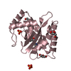
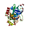
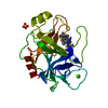
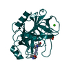
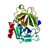
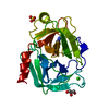
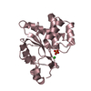
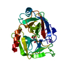
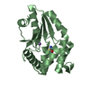
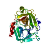
| Title | Crystal structure of Geodia cydonium BHP2 in complex with Lubomirskia baicalensis Bak-2 | ||||||
 Components Components |
| ||||||
 Keywords Keywords | APOPTOSIS / BHP2 / LB-Bak-2 / sponge / Bcl-2 | ||||||
| Function / homology |  Function and homology information Function and homology informationextrinsic apoptotic signaling pathway in absence of ligand / intrinsic apoptotic signaling pathway in response to DNA damage / regulation of apoptotic process / mitochondrial outer membrane / protein heterodimerization activity / apoptotic process / protein homodimerization activity / membrane Similarity search - Function | ||||||
| Biological species |  Geodia cydonium (invertebrata)Lubomirskia baicalensis (Lake Baikal sponge) Geodia cydonium (invertebrata)Lubomirskia baicalensis (Lake Baikal sponge) | ||||||
| Method |  X-RAY DIFFRACTION / SYNCHROTRON / MOLECULAR REPLACEMENT / Resolution: 1.85 Å X-RAY DIFFRACTION / SYNCHROTRON / MOLECULAR REPLACEMENT / Resolution: 1.85 Å | ||||||
 Authors Authors | Caria, S. / Hinds, M.G. / Kvansakul, M. | ||||||
 Citation Citation | Journal: Cell Death Dis / Year: 2017 Title: Structural insight into an evolutionarily ancient programmed cell death regulator - the crystal structure of marine sponge BHP2 bound to LB-Bak-2. Authors: Caria, S. / Hinds, M.G. / Kvansakul, M. | ||||||
| History |
|
- Structure visualization
| Structure viewer | Molecule: MolmilJmol/JSmol |
|---|
- Downloads & links
-Download
| PDBx/mmCIF format | 5twa.cif.gz | 170.2 KB | Display | PDBx/mmCIF format |
|---|---|---|---|---|
| PDB format | pdb5twa.ent.gz | 134.5 KB | Display | PDB format |
| PDBx/mmJSON format | 5twa.json.gz | Tree view | PDBx/mmJSON format | |
| Others |  Other downloads Other downloads |
-Validation report
| Arichive directory | https://data.pdbj.org/pub/pdb/validation_reports/tw/5twaftp://data.pdbj.org/pub/pdb/validation_reports/tw/5twa | HTTPS FTP |
|---|
-Related structure data
| Related structure data | 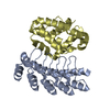 4k5bS S: Starting model for refinement |
|---|---|
| Similar structure data |
-Links
 PDBj
PDBj
- Assembly
| Deposited unit | 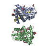
| ||||||||
|---|---|---|---|---|---|---|---|---|---|
| 1 | 
| ||||||||
| 2 | 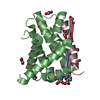
| ||||||||
| Unit cell |
|
-Components
| #1: Protein | Mass: 20864.043 Da / Num. of mol.: 2 / Fragment: UNP residues 19-200 Source method: isolated from a genetically manipulated source Source: (gene. exp.) Geodia cydonium (invertebrata) / Gene: bhp2 / Production host:  #2: Protein/peptide | Mass: 2750.951 Da / Num. of mol.: 2 / Fragment: UNP residues 64-88 / Source method: obtained synthetically Source: (synth.) Lubomirskia baicalensis (Lake Baikal sponge)References: UniProt: Q1RPT5 #3: Chemical | ChemComp-EDO /   Mass: 62.068 Da / Num. of mol.: 20 / Source method: obtained synthetically / Formula: C2H6O2 Mass: 62.068 Da / Num. of mol.: 20 / Source method: obtained synthetically / Formula: C2H6O2#4: Chemical |   Mass: 282.334 Da / Num. of mol.: 2 / Source method: obtained synthetically / Formula: C11H26N2O6 / Comment: pH buffer*YM Mass: 282.334 Da / Num. of mol.: 2 / Source method: obtained synthetically / Formula: C11H26N2O6 / Comment: pH buffer*YM#5: Water | ChemComp-HOH / |  Mass: 18.015 Da / Num. of mol.: 171 / Source method: isolated from a natural source / Formula: H2O Mass: 18.015 Da / Num. of mol.: 171 / Source method: isolated from a natural source / Formula: H2O |
|---|
-Experimental details
-Experiment
| Experiment | Method: X-RAY DIFFRACTION / Number of used crystals: 1 |
|---|
- Sample preparation
| Crystal | Density Matthews: 2 Å3/Da / Density % sol: 38.46 % / Description: Rod crystals |
|---|---|
| Crystal grow | Temperature: 293.15 K / Method: vapor diffusion, sitting drop Details: 28.7% (w/v) PEG 1500, 0.1 M bis-Tris chloride. Protein concentration 5mg/mL |
-Data collection
| Diffraction | Mean temperature: 100 K |
|---|---|
| Diffraction source | Source: SYNCHROTRON / Site: Australian Synchrotron  / Beamline: MX2 / Wavelength: 0.9537 Å / Beamline: MX2 / Wavelength: 0.9537 Å |
| Detector | Type: ADSC QUANTUM 210r / Detector: CCD / Date: Aug 19, 2015 |
| Radiation | Monochromator: double crystal Si(111) / Protocol: SINGLE WAVELENGTH / Monochromatic (M) / Laue (L): M / Scattering type: x-ray |
| Radiation wavelength | Wavelength: 0.9537 Å / Relative weight: 1 |
| Reflection | Resolution: 1.85→41.08 Å / Num. obs: 31937 / % possible obs: 99.7 % / Redundancy: 5 % / Rmerge(I) obs: 0.13 / Net I/σ(I): 8.1 |
| Reflection shell | Resolution: 1.85→1.89 Å / Redundancy: 5 % / Rmerge(I) obs: 1.08 / % possible all: 99.9 |
- Processing
| Software |
| ||||||||||||||||||||||||||||||||||||||||||||||||||||||||||||||||||||||||||||||||||||
|---|---|---|---|---|---|---|---|---|---|---|---|---|---|---|---|---|---|---|---|---|---|---|---|---|---|---|---|---|---|---|---|---|---|---|---|---|---|---|---|---|---|---|---|---|---|---|---|---|---|---|---|---|---|---|---|---|---|---|---|---|---|---|---|---|---|---|---|---|---|---|---|---|---|---|---|---|---|---|---|---|---|---|---|---|---|
| Refinement | Method to determine structure: MOLECULAR REPLACEMENT Starting model: 4K5B Resolution: 1.85→33.947 Å / SU ML: 0.26 / Cross valid method: FREE R-VALUE / σ(F): 1.34 / Phase error: 24.96 / Stereochemistry target values: ML
| ||||||||||||||||||||||||||||||||||||||||||||||||||||||||||||||||||||||||||||||||||||
| Solvent computation | Shrinkage radii: 0.9 Å / VDW probe radii: 1.11 Å / Solvent model: FLAT BULK SOLVENT MODEL | ||||||||||||||||||||||||||||||||||||||||||||||||||||||||||||||||||||||||||||||||||||
| Refinement step | Cycle: LAST / Resolution: 1.85→33.947 Å
| ||||||||||||||||||||||||||||||||||||||||||||||||||||||||||||||||||||||||||||||||||||
| Refine LS restraints |
| ||||||||||||||||||||||||||||||||||||||||||||||||||||||||||||||||||||||||||||||||||||
| LS refinement shell |
| ||||||||||||||||||||||||||||||||||||||||||||||||||||||||||||||||||||||||||||||||||||
| Refinement TLS params. | Method: refined / Origin x: 15.7199 Å / Origin y: -25.3708 Å / Origin z: 0.3265 Å
| ||||||||||||||||||||||||||||||||||||||||||||||||||||||||||||||||||||||||||||||||||||
| Refinement TLS group | Selection details: all |
